
Indications
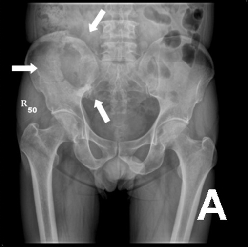
- Mirels score 9 or more
- Over 50% cortical destruction
- Proximal femoral lesion over 2.5 cm or avulsion of the lesser trochanter
- Pain persisting after radiotherapy
- Mirels score uses site, pain, lesion type and size, each scored 1 to 3
- Life expectancy over about 6 weeks for surgery to help
- Myeloma and lymphoma often respond to systemic treatment
Position
- Supine on a traction table, or lateral for arthroplasty
- Image intensifier and long implants available
- Cell salvage is avoided with tumour
Operative Steps
- Biopsy a solitary lesion or unknown primary before fixation. Nailing a primary sarcoma contaminates the whole bone
- Embolise hypervascular renal and thyroid metastases before surgery
- Long cephalomedullary nail to protect the whole bone
- Cement augment or curette the lesion when needed
- Periarticular destruction, cemented long stem arthroplasty or proximal femoral replacement
- Vent the canal before reaming or cementing to reduce embolism
- Send reamings for histology
Postoperative Care
- Weight bear as tolerated
- Radiotherapy to the whole operated field
- Oncology review
- VTE prophylaxis, as cancer raises risk
- Plan bisphosphonate or denosumab with oncology
Complications
- Fat embolism and cardiorespiratory collapse during reaming
- Disease progression and fixation failure
- Bleeding
- Implant failure if the lesion does not respond to treatment
- Infection, higher after radiotherapy
- Wound breakdown
Related Pages
Reviewed by Professor Phong Tran, Head of Orthopaedic Surgery, Western Health. Last updated 10 October 2026.