
Indications
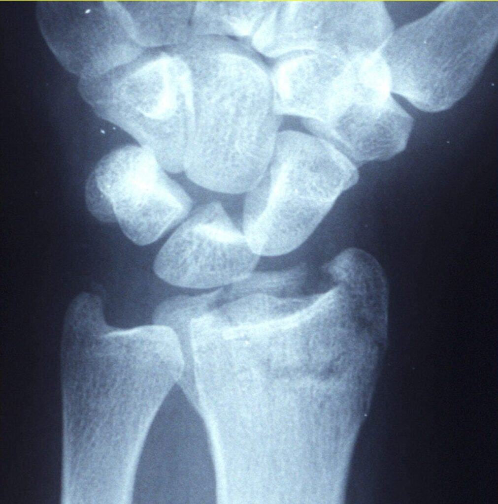
- Displaced fractures that are stable once reduced
- Most paediatric forearm and distal radius fractures
- Temporary splintage before fixation
- Paediatric fractures tolerate more angulation due to remodelling, more so near the physis and in the plane of joint motion
- Rotational deformity does not remodel
Position
- Position for gravity assisted traction where possible
- Analgesia with haematoma block, Bier block or procedural sedation
- Image intensifier to confirm reduction
Operative Steps
- Apply traction to disimpact
- Recreate then reverse the mechanism of injury
- Use the intact soft tissue hinge to hold reduction
- Three point moulding (Charnley)
- Immobilise the joint above and below for shaft fractures
- Pad bony prominences, split or use a backslab if swelling is expected
- Check films in two planes
- Cast index under about 0.8 indicates a good mould in forearm casts
- Avoid excess wrist flexion (Cotton Loder position)
- Convert to fixation if reduction is lost or unstable
Postoperative Care
- Elevation and neurovascular observations
- Repeat X-ray at about 1 week for unstable patterns
- Written cast care advice
- Return if pain worsens, fingers swell or colour changes
- Typical cast duration of 4 to 6 weeks for wrist fractures
Complications
- Compartment syndrome and tight cast
- Pressure areas
- Loss of reduction
- Joint stiffness and CRPS
- Cast saw burns
- Malunion
- Joint stiffness, worse in older patients
Related Pages
Reviewed by Professor Phong Tran, Head of Orthopaedic Surgery, Western Health. Last updated 10 October 2026.