
Indications
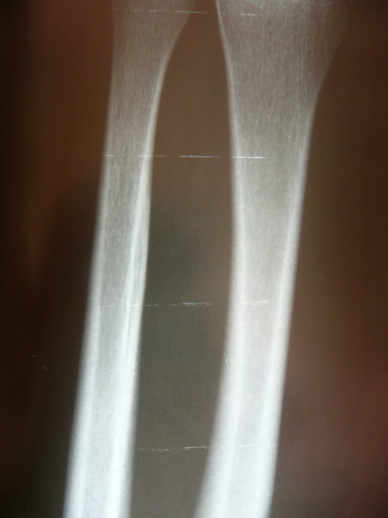
- Displaced both bone fractures in adults
- Monteggia and Galeazzi injuries
- Isolated displaced radial shaft fractures
- Forearm fractures in adults behave like intra-articular injuries because rotation depends on both bones
- Closed treatment of displaced both bone fractures in adults gives poor results
Position
- Supine, arm table, upper arm tourniquet
- Image intensifier with views of the elbow and wrist
Operative Steps
- Radius through the volar Henry approach, supinate proximally to protect the posterior interosseous nerve
- Ulna through the subcutaneous border between ECU and FCU
- Use separate incisions to reduce synostosis risk
- Compression plates, at least three bicortical screws each side
- Restore radial bow for rotation
- Check DRUJ and radiocapitellar joint stability after fixation
- Fix the less comminuted bone first to restore length
- Use 3.5 mm compression plates
- Bone graft is rarely needed with modern biological plating
Postoperative Care
- Early motion
- Avoid plate removal unless symptomatic because of refracture risk
- Sling and light dressing, with motion from the first days
- Radiographs at 2 and 6 weeks
- Return to heavy work after union at about 3 months
Complications
- Radioulnar synostosis
- Posterior interosseous nerve injury
- Refracture after plate removal
- Compartment syndrome
- Malunion with loss of radial bow limits rotation
- Nonunion is uncommon with compression plating
- Infection
Related Pages
Reviewed by Professor Phong Tran, Head of Orthopaedic Surgery, Western Health. Last updated 10 October 2026.