
Indications
- Degenerative spondylolisthesis with stenosis needing decompression
- Isthmic spondylolisthesis with persistent symptoms after conservative care
- Iatrogenic instability after wide decompression or facetectomy
- Degenerative scoliosis, and reconstruction after tumour, infection or fracture
Position
- Prone on a radiolucent Jackson or Wilson frame with the abdomen hanging free
- Free abdomen lowers epidural venous pressure and blood loss
- Hips extended to preserve lumbar lordosis
- Pad eyes, ulnar nerves and anterior superior iliac spines, and confirm fluoroscopic access
Operative Steps
- Localise the level with fluoroscopy and make a midline incision
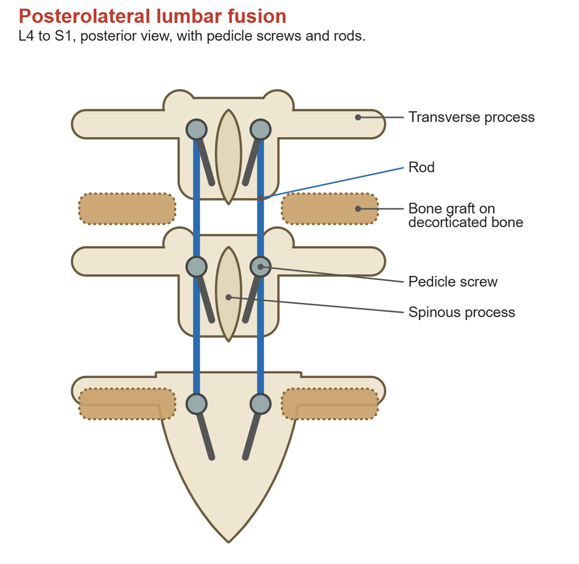
- Dissect subperiosteally along spinous processes and laminae to the facets, preserving the capsule of the cranial adjacent facet
- Continue laterally to the tips of the transverse processes, coagulating segmental vessels at the lateral border
- Entry point lies at the junction of the transverse process midline and the lateral border of the superior facet
- Open cortex with an awl, probe the pedicle and palpate for four intact walls with a ball-tip feeler
- Medial angulation increases caudally and is greatest at L5. Tap, then insert screws under fluoroscopy
- Decorticate transverse processes, pars and lateral facets with a burr until bleeding bone shows
- Pack graft over the decorticated bed bilaterally. Iliac crest autograft has the highest fusion rate but causes donor site pain
- Local laminectomy bone is a standard supplement. Extenders include allograft chips, demineralised bone matrix and ceramics
- BMP-2 is off label in the posterolateral gutter. Risks include ectopic bone, radiculitis, swelling and osteolysis
- Contour rods to lordosis, seat, lock and compress across the segment, then confirm with final imaging
Postoperative Care
- Mobilise on day 1 with physiotherapy and venous thromboembolism prophylaxis
- Smoking cessation supports fusion
- Serial radiographs assess implant position, with CT for suspected non-union
Complications
- Dural tear, repaired primarily with sealant and a period of flat bed rest
- Pseudarthrosis, more likely with smoking, diabetes, osteoporosis and multilevel fusion
- Adjacent segment disease above the construct, worsened by violation of the cranial facet
- Screw malposition, with medial breach threatening the nerve root and anterior S1 breach threatening iliac vessels
- Deep infection and epidural haematoma
Reviewed by Professor Phong Tran, Head of Orthopaedic Surgery, Western Health. Last updated 20 October 2026.