
Indications
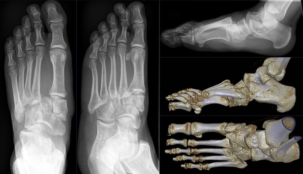
- Unstable tarsometatarsal injuries
- Diastasis between the first and second metatarsal bases on weight bearing or stress views
- Primary partial arthrodesis for purely ligamentous injuries (Ly and Coetzee, JBJS 2006)
- Suspect the injury with plantar arch bruising and midfoot pain after a twisting or crush injury
- Fleck sign at the base of the second metatarsal indicates a ligament avulsion
- CT for fracture pattern and MRI for subtle ligament injury
Position
- Supine, thigh tourniquet
Operative Steps
- Two dorsal longitudinal incisions with wide skin bridges
- Protect the deep peroneal nerve and dorsalis pedis artery
- Reduce the second metatarsal base into its mortise
- Lisfranc screw from the medial cuneiform to the second metatarsal base
- Medial column with transarticular screws or dorsal bridge plates
- Lateral column with K wires to keep it mobile
- Delay surgery until swelling settles, often 10 to 14 days
- Bridge plating avoids articular damage from transarticular screws
- Anatomical reduction is the main determinant of outcome
Postoperative Care
- Non weight bearing for 6 to 8 weeks
- Remove lateral wires at about 6 weeks
- Progressive weight bearing in a boot from about 8 weeks
- Medial column hardware is often removed at about 4 to 6 months unless fused
Complications
- Post-traumatic arthritis
- Foot compartment syndrome
- Neurovascular injury
- Loss of reduction
- Missed injury, about 20% at first presentation
- Hardware failure and painful hardware
Related Pages
Reviewed by Professor Phong Tran, Head of Orthopaedic Surgery, Western Health. Last updated 10 October 2026.