
Anatomy
- Branch of the common peroneal nerve at the fibular neck
- Pierces the anterior intermuscular septum and EDL
- Runs with the anterior tibial artery on the interosseous membrane
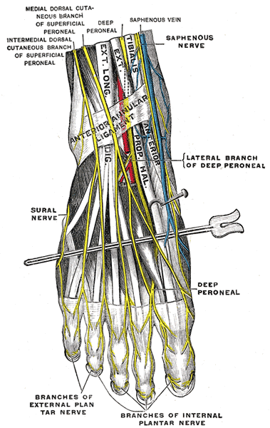
- Crosses the ankle between EHL and EDL, lateral to the dorsalis pedis artery
- Passes beneath the inferior extensor retinaculum and ends in the first web space
- Lateral terminal branch supplies EDB and the tarsal joints
Motor Supply
- Tibialis anterior, EHL, EDL and peroneus tertius
- EDB and EHB
Sensory Supply
- First web space
Clinical Features
- Proximal injury with the common peroneal nerve causes foot drop
- First web space numbness is an early sign of anterior compartment syndrome
- Anterior tarsal tunnel syndrome under the inferior extensor retinaculum follows tight laces, dorsal osteophytes or a ganglion
- Wasting of EDB in chronic distal compression
- Dorsal foot pain worse with plantarflexion and tight shoes
Investigations
- Nerve conduction studies
- Radiographs for dorsal talonavicular osteophytes
Differential Diagnosis
- L5 radiculopathy
- Common peroneal nerve palsy
- Tibialis anterior tendon rupture
Management
- Release of the inferior extensor retinaculum with osteophyte excision
- Tibialis posterior transfer for permanent foot drop
Structures at Risk
- Anterior approach to the ankle
- Anterocentral ankle arthroscopy portal, which is avoided
- Dorsal midfoot approaches and anterior tibial external fixator pins
Reviewed by Professor Phong Tran, Head of Orthopaedic Surgery, Western Health. Last updated 10 October 2026.