
Indications
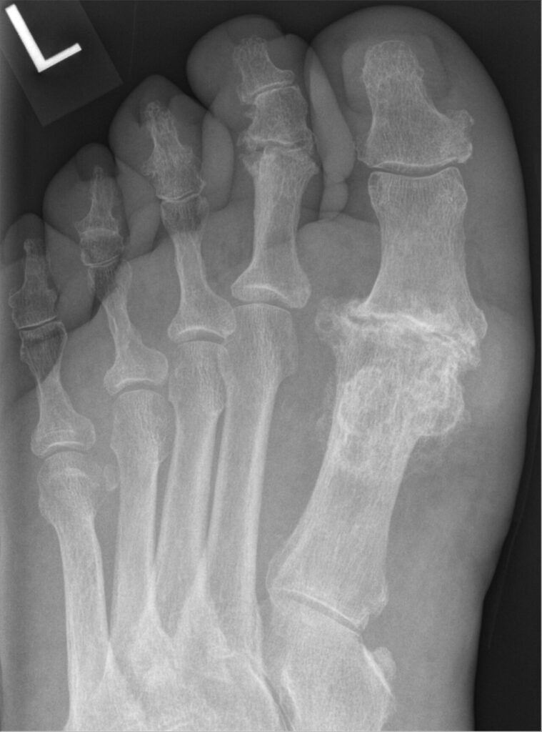
- Advanced hallux rigidus
- Severe or recurrent hallux valgus
- Rheumatoid forefoot
- Salvage of failed surgery
- Neuromuscular disease with hallux deformity
- Hallux varus after previous surgery
Position
- Supine, ankle tourniquet
- Bump under the ipsilateral hip to bring the foot to neutral
- Image intensifier available
Operative Steps
- Dorsal or medial approach, protecting the dorsomedial cutaneous nerve
- Prepare joint surfaces with cup and cone reamers
- Position in about 10 to 15 degrees valgus and neutral rotation
- Dorsiflexion so the pulp just clears a flat plate when loaded
- Fix with a lag screw and dorsal plate
- Release the collateral ligaments and remove osteophytes to deliver the joint
- Drill the subchondral bone to promote fusion
- Hold provisionally with K wires and check position on a flat plate and image intensifier
- Bone graft for defects after failed arthroplasty
Postoperative Care
- Heel weight bearing for about 6 weeks
- Radiographs at 6 and 12 weeks
- Return to normal shoes at about 8 to 10 weeks
- Union rates above 90% with modern fixation
Complications
- Nonunion
- Malposition, too much dorsiflexion causes shoe pressure
- Interphalangeal joint arthritis
- Symptomatic hardware
- Malposition in varus causes pressure on the second toe
- Infection and wound breakdown
- Transfer metatarsalgia
Related Pages
Reviewed by Professor Phong Tran, Head of Orthopaedic Surgery, Western Health. Last updated 10 October 2026.