
Western Health Orthopaedic Registrar presentation – Hallux Valgus Rigidus By Dr Marc Friso
- Bunion
- Derived from latin word Bunio meaning Turnip.
- Consists of:
- 1. Medial eminence
- 2. Bursal sac communicating with joint
- 3. Callosity of skin
- Condition in which there is
- static subluxation if the first MTPJ
- lateral deviation of the great toe
- medial deviation of the first metatarsal
- It is not a simple deformity but complex deformity of the first ray
- Frequently associated with deformity and symptoms of the lesser toes
Epidemiology
- Almost exclusively seen in the shoe wearing population-33% with shoes and 1.9% without.
- Females: Males – 9:1.
Aetiology
- Multifactorial
- Strong hereditary predisposition
- Occasionally only isolated exostosis of the first MTPJ
- Maternal transmission
- hypermobility
- Predisposing factors include;
- Ill fitting stylish shoes
- Where shoes are not worn incidence equal among men & women,
- where shoes are worn, women>men 3:1 to 15:1
- Local Anatomic Factors
- Bone
- Excessive length of first ray
- Excessive rounding of first metatarsal head or incongruous surface
- Obliquity of the first Metatarso- Cuneiform joint
- excessive valgus tilt
- Amputation of second toe
- Pes planus (pronated flatfoot)
- Soft tissue
- Achilles tendon contracture
- limits dorsiflexion and ext rotation of foot occurs with medial force on foot increased.
- Tibialis Posterior – abnormal insertion
- Achilles tendon contracture
- Bone
- General Factors
- Neurogenic imbalance
- ie Cerebral Palsy, Spinal dysraphism, spinal cord tumour
- Rheumatoid Arthritis
- pes valgus with pronated forefoot and increased valgus moment at toe off.
- Generalised ligamentous laxity
- Neurogenic imbalance
- Ill fitting stylish shoes
Anatomy
- Anatomical Features
- Sesamoid bones (within tendons of flexor hallucis brevis, lie in grooves on plantar surface of 1st metatarsal head
- Plantar plate – condensation of FHB, AbH, AdH, plantar fascia
- There are no tendon insertions onto metatarsal head, its position is influenced by position of proximal phalanx
- Base of 1st MTP – articulates with medial cuneiform – may be lax in some cases
Pathogenesis / Pathology
Biomechanics
- Great toe stabilizes the medial foot via the Windlass effect of plantar aponeurosis.
- As body passes over the great toe the PP dorsiflexes and then the Plantar aponeurosis tightens and depresses the first metatarsal
- 50 % of weight through the first metatarsal.
- If the MTPJ subluxes laterally then weight is transferred laterally via the other metatarsal heads
- Must correct this mechanism in surgery for Hallux Valgus
Pathooanatomy
- Stable MTP joints have flatter articulations
- Rounded articulations can progress to hallux valgus deformity
- When articulation is rounded, progressive hallux valgus can develop
- As phalanx deviates laterally, places pressure against metatarsal head, increasing metatarsal – first phalanx angle
- Medial joint structures become attenuated, lateral joint structures contract
- Sesamoids generally remain in place (anchored to adductor hallucis), other structures rotate around joint
- As adductor tendon rotates, becomes more plantar & no longer stabilizes metatarsal head, & becomes deforming force & pronates proximal phalanx
- FHL pulls phalanx laterally
- As deformity progresses, great toe pronates & sesamoid displaces laterally
- Cresta (ridge that stabilizes the sesamoids) is worn down and the lateral sesamoid uncovered
- Final result is hallux can no longer carry its share, & weight is transferred laterally to lesser toe metatarsal heads
- Congruous MTP
- when articular surface of proximal phalanx is parallel to surface of metatarsal head – this is relatively stable
- Incongruous MTP
- occurs when articular surfaces of proximal phalanx & metatarsal head are no longer parallel – this is relatively unstable
Pathophysiology
- HVA < 35
- no pronation of great toe
- HVA > 35
- great toe pronates
- moves abductor hallucis plantarward
- leaving medial capsular ligament as the only restraining medial structure
- there is now no opposition to pull of ADDuctor Hallucis
- which further pulls toe into valgus, further stretching the medial capsule
- FHL, FHB, EHL worsen the valgus pull
Classification
- 1. Adult acquired
- 2. Adolescent
- 3. Rheumatoid
History
- • History to ascertain main complaint.
- − Pain
- Location
- − bunion
- The dorsum -osteophyte
- The plantar aspect from sesamoids
- The inflammed bursa
- Location
- − Determine patient’s main problem → cosmesis, transfer metatarsalgia, second toe deformity, shoe wear, pain
Examination
- Questions to answer
- Severity
- HV angle
- Pronation
- Reducible or fixed
- OA of MTP, IP
- Hypermobile TMT
- Look for callosity under 2nd MT head
- Ankle equinus deformity
- Severity
Shoe Wear/Fit
- Inspect footwear for stretching and bulging over the medial forefoot
- Narrow toe box and high heels load the forefoot and aggravate bunion pain
- Difficulty finding comfortable shoes is a common reason for presentation
- Check sole wear pattern for altered weight bearing and lateral forefoot overload
- Compare shoe width with weight bearing forefoot width
Look
- Standing
- Front
- General alignment
- Hallux valgus
- Degree
- pronation
- Bunion
- Lesser toe abnormalities
- skin
- Side
- Pes planus
- Longitudinal arch
- Behind
- Foot in standing posture
- Posture of forefoot in relation to hindfoot
- Front
- GAIT
- Sitting
- Dorsum
- Medial eminence – redness/ulceration
- Ingrown toenail
- Plantar
- Callosities under metatarsal heads
- Dorsum
Feel
- Tenderness
- Dorsal osteophyte 1st MTPJ
- tenderness over medial aspect of MTP joint
- occurs from irritation of dorsal cutaneous nerve
- may occur from hypertrophic bursa
Move
- Hallux valgus reducible
- ROM
- TA tightness
- ankle,
- subtalar,
- midtarsal,
- TMTJ
- Pain
- Laxity / stable
- ROM
- MTPJ
- Reducible
- Irritable
- crepitus, loss of motion and pain
- MTPJ
- IPJ
- Quantify range
- ? irritable
Special
- Neurovascular status
- Sensation
- Medial / lateral side of 1st toe
- Medial side of 2nd toe
- Ligamentous laxity
Investigations
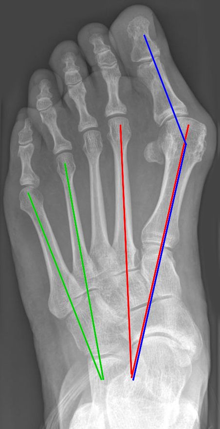
Standing Radiographs (AP, lat, oblique views)
- Angles
| Angle | Normal | Importance |
|---|---|---|
| HVA (hallux valgus angle) | <15 | |
| IMT (Intermetatarsal angle) | <9 | |
| DMMA (Distal metatarsal articular angle) | <10° | |
| Hallux Valgus Interphalangeus | < 10° | |
| Metatarsus Primus Varus | < 25 |
- High DMMA = congruent joint = osteotomy needed not lateral release
- Congruency of first MTPJ
- Noncongruency = lateral release
- Sesamoid congruency
- determined relative to a line that bisects the long axis of the first metatarsal shaft
| Grade | Description |
|---|---|
| 1 | < 50% overlap the reference line |
| 2 | > 50% overlap the reference line |
| 3 | complete displacement beyond reference line |
- Size of medial eminence
- First MTPJ OA
- IPJ OA
- Obliquity of Metatarsocuneiform joint
- Hypermobility signs
- Plantar gapping
- 2nd MT cortical thickening
Treatment
Primary Goals
- Reduce pain
- Restore articular congruency of first ray
- Restore alignment of first ray relative to the lateral part of foot
- Patients shoe wear habits essential
- Before surgery only 1/3 of patients wear shoes they want and after surgery only 2/3 are able to wear shoes of choice.
- Surgery should not be done in asymptomatic feet.
- Will fail if predisposing factors not addressed as well.
Need to Consider
- Occupation and athletic pursuits
- Patient expectations
- Failure of non-op measures
- Age
- Physical findings
- Radiographic abnormalities
- Neurovascular status
Non-operative
- ELPOMI
- Wide toe box and broad shoe
- Low heels
- Avoid seams
- Soft expandable upper
- Premetatarsal bar and medial arch support
- Podiatrist for callosities.
- Bunion splint
- Toe spacer
- Orthosis with medial arch
- Shoe stretching
Operative
- Indications
- Pain
- Shoewear difficulties
- Ulceration (neuropathic)
- HVA > 20°
- IMA > 10°
- Failed nonoperative management
- Contraindications
- Cosmetic
- Active Athlete
- Unrealistic expectations
- vascular insufficiency
- neuropathic foot
- Children with open physis
- Spastic muscular condition
- Severely pronated foot (high rate of recurrence)
- Ideal hallux valgus surgery
- 1. Correction of the IMT and HV angles
- a. Minimal shortening
- b. Adequate stability
- 2. Creation of a congruent 1st MTPJ with sesamoid realignment
- 3. Resection of the medial eminence parallel to and flush with the MT shaft
- 4. Retention of function and ROM of the 1st MTPJ
- 5. Maintenance of normal weight bearing mechanics
- 1. Correction of the IMT and HV angles
Algorithm
| Severity | Congruent | Noncongruent |
|---|---|---|
| Mild HV < 20 IMT <10 | * Chevron osteotomy * Mitchell’s osteotomy | * Distal soft tissue procedure ~ (Adductor Hallucis, intermetatarsal lig. & lateral capsule) * Chevron osteotomy * Mitchell’s osteotomy |
| Moderate HV <40 IMT <20 | Scarf + Akin + Distal soft tissue procedure | Scarf + Akin + Distal soft tissue procedure |
| Severe HV >40 IMT >20 | Scarf + Akin + Distal soft tissue procedure | Scarf + Akin + Distal soft tissue procedure |
- OA = arthrodesis
- Fusion
- Hypermobile 1st MTPcuneiform
- this joint should be fused, & distal soft tissue procedure performed.
- Keller in low demand elderly pt
- Juvenile Hallux Valgus
- Delay surgery until skeletal maturity
- ↑ DMAA
- ↑ recurrence
Complications
- Deformity
- Overcorrection
- Hallux Varus
- Negative IM angle
- Excess MT head resection
- Lateral sesamoid excision
- Recurrence
- Failure to include MT osteotomy
- Inadequate lateral release
- Poor quality medial tissue
- Cock up deformity
- Pain
- Transfer metatarsalgia
- Shortening
- Dorsiflexion
- Stiffness / OA
- AVN
- 0 to 20% for chevron, 8 to 12% for Mitchell
Bunion Procedures
1. Distal Soft Tissue Procedures.
- Modified McBride
2. Distal Bony Procedures.
- Chevron Osteotomy
- Mitchell Osteotomy
- Wilsons Shaft Osteotomy
3. Proximal Metatarsal Osteotomy
- Scarf
4. Proximal Phalangeal Osteotomy.
- Akin Procedure
5. Kellers Resection Arthroplasty.
6. Arthrodesis of 1st MTPJ
7. Arthroplasty of 1st MTPJ
8. Metatarsocunieform Arthrodesis (1st TMT Lapidus)
Reviewed by Professor Phong Tran, Head of Orthopaedic Surgery, Western Health. Last updated 10 October 2026.