
Indications
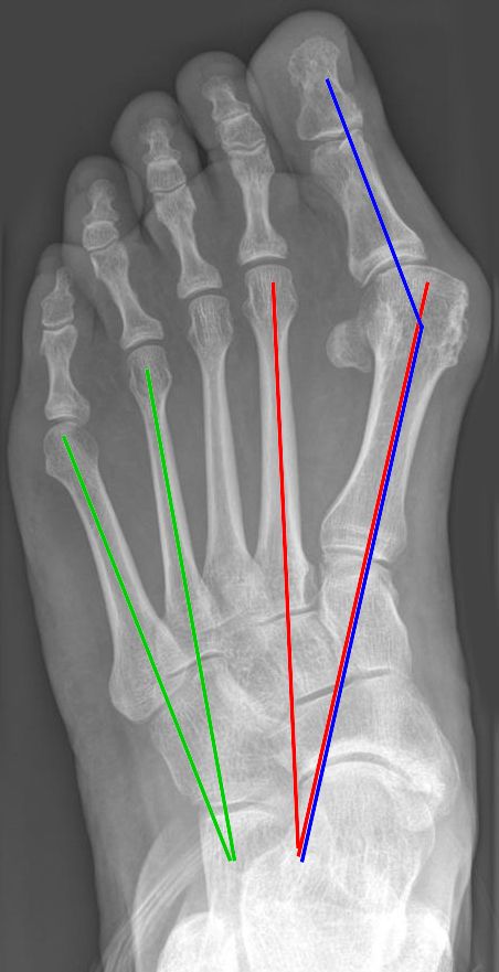
- Painful bunion and shoe wear problems despite footwear changes
- Surgery is not offered for cosmetic reasons alone
- Assess for first ray hypermobility, lesser toe deformity and transfer metatarsalgia
Position
- Supine, ankle or thigh tourniquet
- Bump under the ipsilateral hip
- Image intensifier available
Operative Steps
- Measure hallux valgus angle, intermetatarsal angle and distal metatarsal articular angle on weight bearing films
- Mild to moderate deformity, distal chevron osteotomy
- Moderate to severe deformity, scarf or proximal osteotomy
- Hypermobile first ray or severe deformity, Lapidus first TMT fusion
- Akin osteotomy for residual interphalangeal valgus
- Lateral release of adductor hallucis and capsule, preserving the fibular sesamoid
- Medial capsulorrhaphy
- Normal values are HVA under 15 degrees and IMA under 9 degrees
- Minimally invasive distal osteotomy is an alternative in selected patients
- Check sesamoid position and joint congruency after correction
Postoperative Care
- Heel weight bearing in a postoperative shoe for about 6 weeks
- Radiographs at 6 weeks
- Return to normal shoes at about 8 to 12 weeks
- Swelling can last 6 to 12 months
Complications
- Recurrence
- Hallux varus from over release
- Avascular necrosis of the metatarsal head
- Transfer metatarsalgia and stiffness
- Malunion with dorsiflexion of the first metatarsal
- Nonunion, most often after Lapidus fusion
- Dorsomedial cutaneous nerve injury
Related Pages
Reviewed by Professor Phong Tran, Head of Orthopaedic Surgery, Western Health. Last updated 10 October 2026.