
Indications
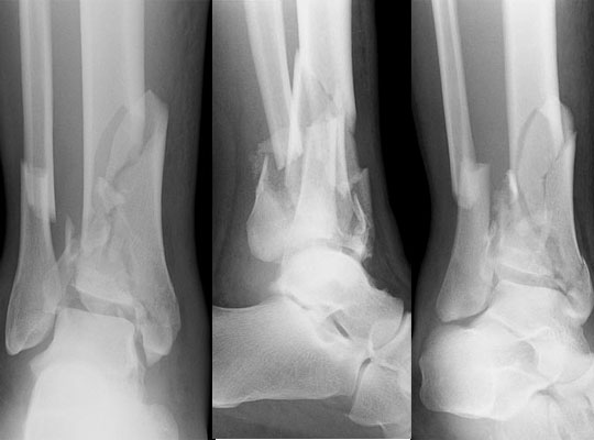
- Displaced intra-articular distal tibial fractures
- High energy axial loading injuries with soft tissue compromise
- Tscherne grading guides timing of definitive surgery
- Open fractures need urgent debridement and stabilisation
Position
- Supine, bump under the hip, thigh tourniquet
Operative Steps
- Stage one, spanning external fixation and fibular fixation if the skin allows
- CT with the frame on to plan approaches
- Stage two at about 10 to 21 days once the skin wrinkles
- Anteromedial, anterolateral or posterolateral approaches chosen from the fracture lines, with wide skin bridges
- Reconstruct the articular surface, then attach it to the shaft
- Graft metaphyseal defects and buttress the medial or anterior column
- Rüedi and Allgöwer principles, fibular length, articular reduction, bone graft, buttress plate
- Ring fixators with limited internal fixation are an alternative in poor soft tissue
- Primary ankle fusion in severe comminution in older patients
Postoperative Care
- Elevation and wound checks
- Non weight bearing for 8 to 12 weeks
- Early ankle and subtalar motion when the wound allows
- Radiographs at 2, 6 and 12 weeks
- Return to work can take many months
Complications
- Wound breakdown and deep infection
- Post-traumatic arthritis
- Malunion and nonunion
- Stiffness of the ankle
- Late fusion for arthritis in some patients
- Complex regional pain syndrome
Related Pages
Reviewed by Professor Phong Tran, Head of Orthopaedic Surgery, Western Health. Last updated 10 October 2026.