
Indications
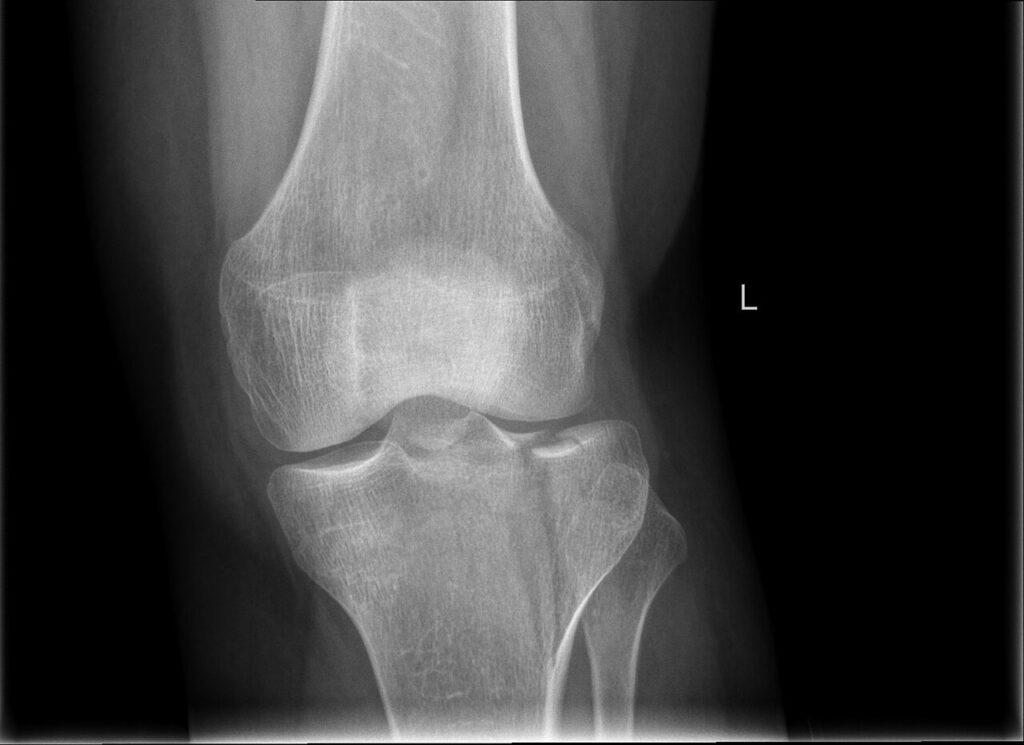
- Articular step or condylar widening
- Malalignment and instability
- Spanning external fixation first when soft tissues are poor
- Schatzker classification and CT based three column concepts guide fixation
- Articular step over about 2 mm and condylar widening over 5 mm are common thresholds
- Undisplaced fractures are treated in a hinged brace
Position
- Supine, radiolucent table, bump under the hip
- Image intensifier from the opposite side
- Thigh tourniquet available
Operative Steps
- CT to plan approaches and fragment fixation
- Anterolateral approach with submeniscal arthrotomy for lateral fractures
- Elevate depressed articular fragments through a cortical window
- Fill the void with graft or substitute, support with rafter screws
- Lateral buttress or locking plate
- Posteromedial approach for medial or bicondylar fractures, with dual plating
- Avoid a single midline incision with wide stripping
- Repair the meniscus
- Check the lateral meniscus, which is often trapped in the fracture
- Restore the mechanical axis and joint line height
- Hybrid or ring frames for comminuted bicondylar fractures with poor skin
Postoperative Care
- Early motion, protected weight bearing for 6 to 12 weeks
- Radiographs at 2, 6 and 12 weeks
- CPM is not essential if active motion starts early
Complications
- Wound breakdown and deep infection
- Compartment syndrome
- Loss of reduction and post-traumatic arthritis
- Common peroneal nerve injury
- Knee stiffness
- Peroneal nerve injury with lateral approaches
- Malalignment
Related Pages
Reviewed by Professor Phong Tran, Head of Orthopaedic Surgery, Western Health. Last updated 10 October 2026.