
Indications
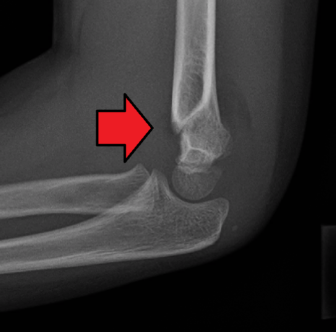
- Displaced Gartland type II and III extension fractures
- Flexion type and unstable fractures
- Pulseless limbs need urgent reduction in theatre
Position
- Supine, arm on a hand table or on the image intensifier
- Image intensifier as the operating table supports the arm
Operative Steps
- Longitudinal traction with the elbow extended
- Correct coronal translation and rotation
- Flex the elbow while pushing the olecranon forward, pronate for posteromedial displacement
- Check Baumann angle and the anterior humeral line through the capitellum
- Two or three divergent lateral pins engaging both columns
- Medial pin only through a small incision with the elbow extended to protect the ulnar nerve
- Assess hand perfusion after reduction
- Avoid excessive flexion in a swollen elbow
- Test stability under fluoroscopy before final splinting
Postoperative Care
- Backslab at about 60 to 90 degrees flexion
- Pins out at 3 to 4 weeks
- Pulseless pink hand, observe closely. Pulseless white hand, explore the brachial artery
- Neurovascular observations and elevation
- Radiographs at 1 week
Complications
- Cubitus varus from malreduction
- Anterior interosseous nerve palsy, the most common nerve injury
- Iatrogenic ulnar nerve injury from medial pins
- Compartment syndrome and pin infection
- Anterior interosseous nerve palsy is the most common nerve injury, usually recovering
- Compartment syndrome
- Pin tract infection
Related Pages
Reviewed by Professor Phong Tran, Head of Orthopaedic Surgery, Western Health. Last updated 10 October 2026.