
Definition
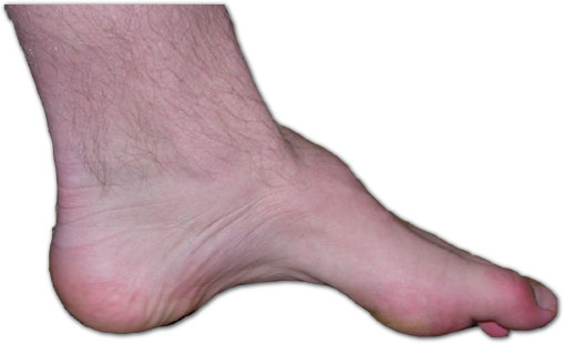
- High medial longitudinal arch from a forefoot plantarflexed on the hindfoot
- Usually cavovarus with hindfoot varus
Aetiology
- Neurological cause in about two thirds
- CMT the most common cause
- Spinal dysraphism, tethered cord, spinal cord tumour and Friedreich ataxia
- Polio and CP
- Residual clubfoot, trauma and compartment syndrome
Pathology
- Weak tibialis anterior is overpowered by peroneus longus, plantarflexing the first ray
- Weak peroneus brevis is overpowered by tibialis posterior, driving hindfoot varus
- Intrinsic weakness produces claw toes
- Plantarflexed first ray forces the hindfoot into varus by the tripod effect
Clinical Features
- Peek a boo heel sign from the front
- Claw toes and metatarsal head callus
- Lateral ankle instability and fifth metatarsal stress fractures
- Unilateral cavus warrants spinal MRI
- Family history, full neurological examination and Romberg test
Investigations
- Coleman block test shows whether hindfoot varus is forefoot driven and flexible
- Standing lateral radiograph with Meary angle apex dorsal
- Increased calcaneal pitch in calcaneocavus
- MRI spine, nerve conduction and genetic testing
Management
- Orthoses with lateral forefoot post for flexible deformity
- Plantar fascia release
- Dorsiflexion osteotomy of the first metatarsal
- Peroneus longus to brevis transfer
- Tibialis posterior transfer through the interosseous membrane for foot drop
- Lateralising calcaneal osteotomy (Dwyer) for fixed hindfoot varus
- Jones transfer of EHL to first metatarsal neck with hallux IP fusion
- Triple arthrodesis as salvage, avoided in young patients
Prognosis
- Progressive in neurological causes
- Joint sparing surgery before deformity becomes rigid gives better results
Reviewed by Professor Phong Tran, Head of Orthopaedic Surgery, Western Health. Last updated 10 October 2026.