
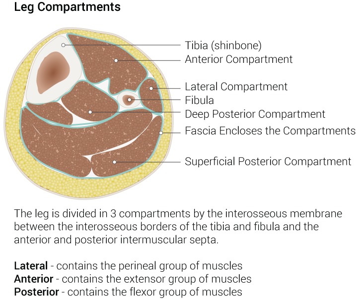
Compartments
| Compartment | Muscles | Nerve | Artery |
|---|---|---|---|
| Anterior | Tibialis anterior, EHL, EDL, peroneus tertius | Deep peroneal | Anterior tibial |
| Lateral | Peroneus longus and brevis | Superficial peroneal | Branches of peroneal |
| Superficial posterior | Gastrocnemius, soleus, plantaris | Tibial (motor branches) | Sural arteries |
| Deep posterior | Tibialis posterior, FDL, FHL, popliteus | Tibial | Posterior tibial and peroneal |
Osteology
- Tibia carries about 85% of the axial load
- Fibula gives attachment to muscles and forms the lateral malleolus
- Nutrient artery of the tibia enters posteriorly in the proximal third and is lost in displaced fractures
Clinical Relevance
- Anterior compartment is most often affected in compartment syndrome
- Two incision fasciotomy decompresses all four compartments. See Compartment Syndrome
- Superficial peroneal nerve pierces the deep fascia about 12 cm above the lateral malleolus
- Behind the medial malleolus, front to back, lie tibialis posterior, FDL, posterior tibial artery, tibial nerve and FHL
- Common peroneal nerve winds around the fibular neck, at risk from plaster, traction and lateral approaches
- Saphenous nerve and great saphenous vein lie anterior to the medial malleolus
- Anterior tibial artery pierces the interosseous membrane and becomes dorsalis pedis at the ankle
- Sural nerve runs with the small saphenous vein behind the lateral malleolus
- Medial face of the tibia is subcutaneous, making open fractures common
- Interosseous membrane fibres run downward and laterally from tibia to fibula
- Deep peroneal nerve supplies the first web space, a quick test after injury
Reviewed by Professor Phong Tran, Head of Orthopaedic Surgery, Western Health. Last updated 10 October 2026.