
Overview
- Commonest inherited peripheral neuropathy
- Leading cause of paediatric cavovarus foot
- Weak tibialis anterior and peroneus brevis against stronger peroneus longus and tibialis posterior
- Result is a plantarflexed first ray, hindfoot varus and claw toes
- Every cavus foot, especially unilateral, needs neurological examination and consideration of spine MRI
- Soft tissue release, tendon transfer and osteotomy, with arthrodesis for rigid deformity
Paediatric Cavus Foot Deformities from Orthofracs on Vimeo.
Presentation from Victorian Tasmanian Orthopaedic Training Program
Aetiology
- Commonest inherited neuropathy and commonest neurological cause of paediatric cavovarus
- CMT1A is a PMP22 duplication on chromosome 17, autosomal dominant, with slow conduction
- CMT2 is axonal with preserved velocities
- X-linked forms involve connexin 32
Pathology
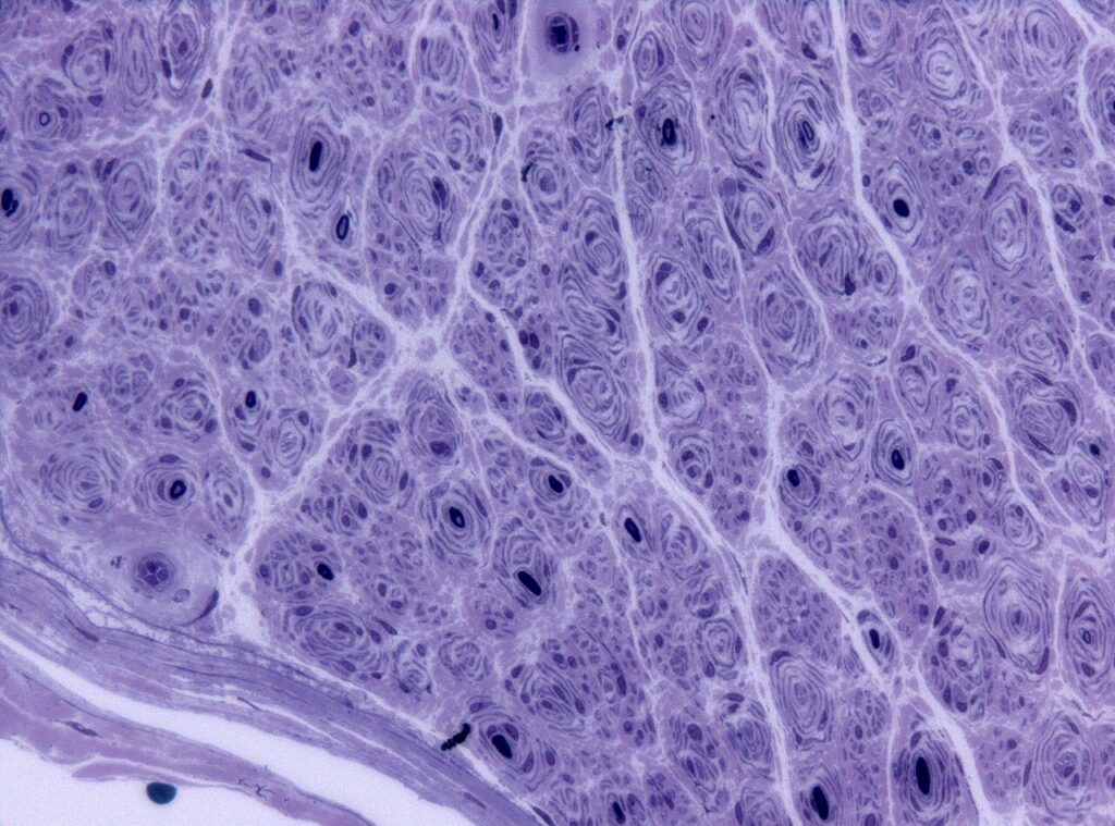
- Weak tibialis anterior is overpowered by peroneus longus, plantarflexing the first ray
- Weak peroneus brevis is overpowered by tibialis posterior, causing hindfoot varus
- Intrinsic weakness causes toe clawing and a high arch
- Forefoot-driven varus is flexible initially, then becomes fixed
Clinical Features
- Peek-a-boo heel sign
- Lateral foot pain, ankle sprains and callosities under first and fifth metatarsal heads
- Steppage gait, absent reflexes and calf and hand wasting
Investigations
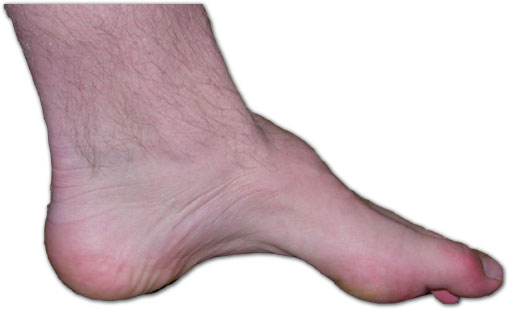
- Coleman block test, hindfoot correction indicates flexible forefoot-driven varus
- Spine MRI if asymmetric or rapidly progressive
- Lateral radiograph shows increased calcaneal pitch and Meary angle
Management
- Bracing does not prevent progression
- Plantar fascia release and first metatarsal dorsiflexion osteotomy
- Peroneus longus to brevis transfer
- Tibialis posterior transfer through the interosseous membrane for foot drop
- Jones procedure for the clawed hallux
- Dwyer lateralising calcaneal osteotomy for fixed hindfoot varus
- Triple arthrodesis is salvage for rigid deformity in the mature foot
Prognosis
- Recurrence is common as the neuropathy progresses
- Joint-sparing reconstruction with tendon balancing gives the best long-term results
Reviewed by Professor Phong Tran, Head of Orthopaedic Surgery, Western Health. Last updated 10 October 2026.