
Definition
- Osteochondrosis of the tarsal navicular with sclerosis, fragmentation and collapse
- Self limiting with reconstitution of the navicular
Aetiology
- Unknown
- Navicular is the last tarsal bone to ossify
- Late ossifying centre is compressed between talus and cuneiforms during weight bearing
- Compression of a precarious blood supply is the favoured theory
Epidemiology
- Children under 6 years
- Boys affected more than girls
- Occasionally bilateral
Clinical Features
- Midfoot pain over the navicular, worse with weight bearing
- Limp, often walking on the lateral border of the foot
- Tenderness, sometimes swelling and warmth over the dorsomedial midfoot
Investigations
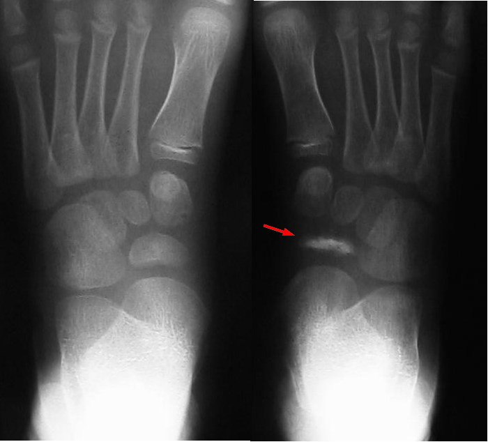
- Radiographs show increased density, fragmentation and narrowing of the navicular
- Flattened navicular reconstitutes over time
- Irregular ossification in an asymptomatic child is a normal variant and not Kohler’s disease
- Compare with the contralateral foot
Differential Diagnosis
- Normal variant of navicular ossification
- Accessory navicular
- Stress fracture
- Osteomyelitis and septic arthritis
- Tarsal coalition in older children
Management
- Symptomatic treatment only
- Activity modification, analgesia and arch support for mild symptoms
- Short leg walking cast for several weeks for marked pain
- Casting shortens the duration of symptoms
- No role for surgery
Prognosis
- Excellent, with radiographic reconstitution over months to years
- No long term disability or deformity reported
Reviewed by Professor Phong Tran, Head of Orthopaedic Surgery, Western Health. Last updated 10 October 2026.