
Indications
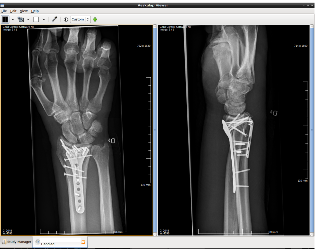
- Unstable or displaced intra-articular fractures
- Loss of reduction in plaster
- Volar shear (Smith and Barton) fractures
- Acceptable alignment after closed reduction is radial inclination over 15 degrees, radial shortening under 3 mm and dorsal tilt under 10 degrees
- Older low demand patients do well in plaster despite malunion
Position
- Supine, arm table, upper arm tourniquet
- Image intensifier and hand table
- Supinate the forearm
Operative Steps
- Modified Henry approach through the FCR sheath
- Retract FPL ulnarly and release pronator quadratus from the radius
- Reduce the fracture, using the brachioradialis release if needed
- Plate proximal to the watershed line to avoid flexor tendon irritation
- Distal screws short of the dorsal cortex to protect extensor tendons
- Check DRUJ stability after fixation
- Use the plate as a buttress for volar shear fractures
- Check the joint with a tilted lateral view
- Dorsal or fragment specific fixation for dorsal shear or die punch fragments
Postoperative Care
- Early finger and wrist motion
- Splint for comfort
- Hand therapy for stiffness
- Return to heavy work at about 3 months
Complications
- FPL rupture from a prominent plate
- Extensor tendon rupture from long screws
- Median nerve symptoms and CRPS
- Loss of reduction
- Malunion with ulnar impaction
- Infection is uncommon
Related Pages
Reviewed by Professor Phong Tran, Head of Orthopaedic Surgery, Western Health. Last updated 10 October 2026.