
Anatomy
- Deep motor branch of the radial nerve, arising at the level of the radiocapitellar joint
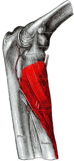
- Passes beneath the arcade of Frohse at the proximal edge of supinator
- Winds around the radial neck within supinator
- Terminal sensory branch lies in the floor of the fourth extensor compartment
Motor Supply
- ECRB in some patients, supinator, ECU, EDC, EDM
- APL, EPL, EPB and EIP
- ECRL and brachioradialis are supplied by the radial nerve before it divides
Clinical Features
- Finger and thumb drop with preserved wrist extension
- Wrist deviates radially on extension because ECRL is spared and ECU is weak
- No sensory loss
- Causes include Monteggia injuries, radial head and neck fractures, lipoma, ganglion and rheumatoid synovitis
- Radial tunnel syndrome gives lateral forearm pain without weakness, with tenderness distal to the lateral epicondyle
Investigations
- MRI or ultrasound for a mass at the radial neck
Management
- Observe closed injuries
- Excise a compressing mass
- Tendon transfers for permanent palsy, such as FCR to EDC and palmaris longus to EPL
Structures at Risk
- Kocher approach to the radial head, so keep the forearm pronated and stay proximal to the annular ligament
- Thompson approach to the proximal radius
- Bicortical drilling of the radial tuberosity in distal biceps repair
Reviewed by Professor Phong Tran, Head of Orthopaedic Surgery, Western Health. Last updated 10 October 2026.