
Anatomy
- Terminal branch of the posterior cord of the brachial plexus, root values C5 and C6
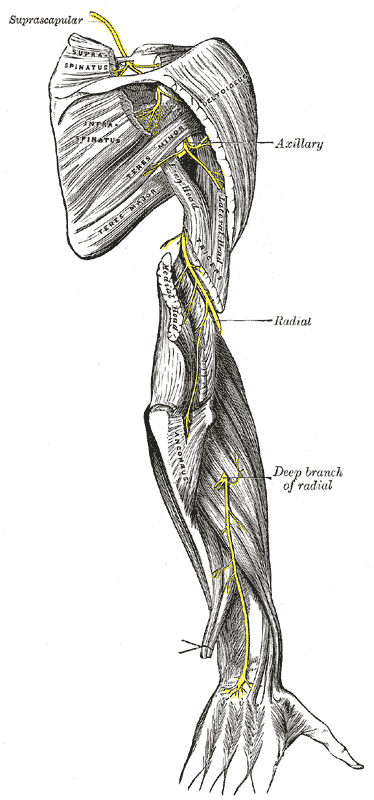
- Passes anterior to subscapularis, then runs close to the inferior shoulder capsule
- Leaves the axilla through the quadrangular space with the posterior circumflex humeral artery
- Anterior branch winds around the surgical neck on the deep surface of deltoid, about 5 cm below the acromion
- Posterior branch supplies teres minor and posterior deltoid, then becomes the superior lateral cutaneous nerve of the arm
Motor Supply
- Deltoid
- Teres minor
Sensory Supply
- Skin over the lower deltoid (regimental badge area)
Clinical Features
- Injured with anterior shoulder dislocation and proximal humerus fractures, more often in older patients
- Weak abduction and loss of deltoid contour
- Sensation can be preserved despite complete motor loss, so test deltoid power
- Quadrilateral space syndrome compresses the nerve in throwing athletes
Investigations
- Nerve conduction studies and EMG at 3 to 4 weeks give a baseline
- MRI for a paralabral cyst or mass in quadrilateral space syndrome
Management
- Most traction injuries after dislocation recover
- EMG if there is no clinical recovery by 3 months
- Nerve grafting or a triceps branch radial to axillary nerve transfer when recovery fails by 6 months
Structures at Risk
- Deltoid splitting approaches extended more than 5 cm below the acromion
- Inferior capsular shift and low posterior arthroscopic portals
- Inferior glenoid during capsular release
Reviewed by Professor Phong Tran, Head of Orthopaedic Surgery, Western Health. Last updated 10 October 2026.