
Anatomy
- Formed from L4, L5 and S1
- Leaves the pelvis through the greater sciatic notch above piriformis with the superior gluteal vessels
- Runs between gluteus medius and gluteus minimus
- Lowest branch lies about 5 cm above the tip of the greater trochanter
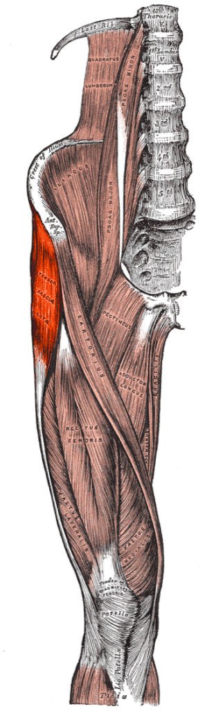
- Superior branch supplies gluteus medius and minimus and the inferior branch continues to tensor fascia lata
Motor Supply
- Gluteus medius
- Gluteus minimus
- Tensor fascia lata
Clinical Features
- Trendelenburg gait and a positive Trendelenburg test
- Abductor weakness after the direct lateral approach to the hip
- Injured with fractures extending into the greater sciatic notch
- Compensated gait with trunk lean over the affected hip
- A persistent limp after hip arthroplasty raises suspicion of injury
Investigations
- EMG of gluteus medius
- MRI to separate denervation from abductor tendon tears
Differential Diagnosis
- Gluteus medius tendon tear
- L5 radiculopathy
Management
- Prevention, as recovery after division is poor
- Gait retraining for partial injuries
- Abductor strengthening and a walking stick in the opposite hand
Structures at Risk
- Splitting gluteus medius more than 5 cm above the greater trochanter in the direct lateral (Hardinge) approach
- Greater sciatic notch dissection in posterior pelvic and acetabular surgery
- Proximal femoral nail insertion
- Anterolateral (Watson-Jones) approach when gluteus medius is retracted forcefully
Reviewed by Professor Phong Tran, Head of Orthopaedic Surgery, Western Health. Last updated 10 October 2026.