
Western Health Orthopaedic Registrar presentation – Proximal Humerus Fractures By Dr James Churchill
Incidence
- Around 4-5% of all fractures
- 85% have minimal displacement – Neer group I
- 5% – severe – 3 or 4 part fractures or fracture dislocations
Anatomy
- average head-humeral shaft angle: 140 degrees
- retroversion: 28.8 degrees
- Displacing Forces:
- Greater tuberosity fracture fragments migrate superiorlyand posteriorly, following the vector of the superior and posterior rotator cuff.
- The lesser tuberosity translates anteriorly and medially along the vector of the subscapularis.
- The superior and posterior rotator cuff is responsible for varus malalignment of the proximal humerus
- The pectoralis major tendon can adduct and translate the humeral shaft anteriorly.
- Landmarks
- The superior margin of the pectoralis major tendon inserts approximately 5.6 cm distal to the top of the humeral head or 4.2 cm distal to the superomedial corner of the greater tuberosity.
- Blood Supply
- anterior circumflex humeral artery and its terminal branch, the arcuate artery, are the main source of perfusion
to the proximal humerus. - significant intraosseous anastomoses between
- the arcuate artery
- posterior circumflex humeral artery,
- metaphyseal arteries,
- vessels of the greater and lesser tuberosities.
- anterior circumflex humeral artery and its terminal branch, the arcuate artery, are the main source of perfusion
Classification
Neer Classification
- Classification is based on the four-part anatomy of the proximal humerus
- humeral head
- lesser tuberosity
- greater tuberosity
- proximal humeral shaft
- Criterion for displacement
- greater than 1 cm of separation of part
- angulation of 45 degrees.
- Neer’s terminology of four-segment classification of displaced fractures and fracture-dislocations relates pattern of displacement (two-part, three-part, or four-part) and key segment displaced.
| Pattern | Description |
|---|---|
| Two-part | In each two-part pattern, segment named is one displaced. Surgical neck fractures may be impacted (A), unimpacted (B) or comminuted (C) |
| Three-part | All three-part patterns have displacement of shaft segment. Displaced tuberosity identifies type of three-part fracture |
| Four-part | All segments are displaced |
| Fracture-dislocation | Identified by anterior or posterior position of articular segment |
Original publication Neer Cs 2nd. Displaced proximal humeral fractures. I. Classification and evaluation. J Bone Joint Surg Am. 1970;52(6):1077-89.
- Large articular surface defects require separate recognition
- Displaced three-part and four-part fractures
- markedly alter the articular congruity of the glenohumeral joint
- highest likelihood of disrupting the major blood supply to the proximal humerus
- Osteonecrosis is most likely after displaced four-part fractures.
- Significant intra & inter-observer variability
Investigations
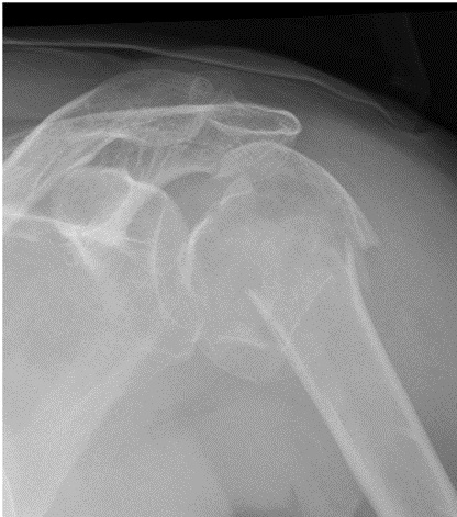
X-rays
- AP, Lateral
CT
- defining head splitting fractures
MRI
- assess for related cuff or labral lesions
- labral lesions are common, occurring in 56% of patients with fracture dislocations.
Treatment
Treatment of Displaced Fractures
Decision based on:
- Age & activity level of patient
- Quality of bone
- Type of fracture
- Presence of other injuries
- Humeral head salvage should be attempted in young patients with good bone stock
- In older patients primary arthroplasty may be performed
Displaced Surgical Neck Fractures
- In adults these should be stabilised after closed reduction with terminally threaded K wires
Tuberosity Fractures
- If these are displaced more than 5mm they should be openly reduced
- greater tuberosity via deltoid splitting approach
- lesser tuberosity via deltopectoral approach
- If GT fractures are not fixed they may cause impingement against acromion
4 Part Fractures
- In young adults, ORIF associated with AVN in around 10%
- In patients treated with hemiarthroplasty, there is usually good pain relief but poor ROM.
- Functional outcomes are better after early hemiarthroplasty (within 4 weeks of fracture) than after late arthroplasty following unsuccessful non-operative treatment.
Reviewed by Professor Phong Tran, Head of Orthopaedic Surgery, Western Health. Last updated 10 October 2026.