
Anatomy
- Formed from L5, S1 and S2
- Leaves the pelvis through the greater sciatic notch below piriformis
- Enters the deep surface of gluteus maximus
- Runs with the inferior gluteal vessels
- Lies close to the sciatic and posterior femoral cutaneous nerves below piriformis
- Supplies no skin
- The posterior femoral cutaneous nerve can share a common trunk with it
Motor Supply
- Gluteus maximus
Clinical Features
- Weak hip extension
- Difficulty climbing stairs and rising from a chair
- Rarely injured in isolation
- Gluteal wasting with flattening of the buttock
- Injured with posterior hip dislocation and posterior acetabular fractures
- Gluteus maximus gait with a backward lurch of the trunk at heel strike
Investigations
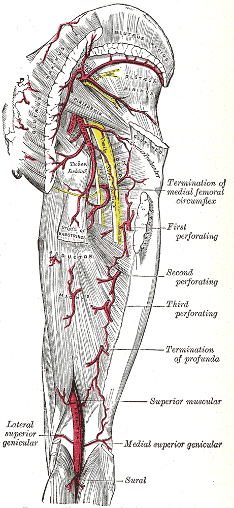
- EMG of gluteus maximus
- MRI of the pelvis for a mass at the greater sciatic notch
- Nerve conduction studies of the sciatic nerve to exclude a combined lesion
Differential Diagnosis
- S1 radiculopathy
- Sciatic nerve injury, which also causes hamstring and foot weakness
Management
- Prevention during posterior approaches
- Physiotherapy with hip extensor strengthening
- No reliable reconstruction exists for complete division
Structures at Risk
- Extending the gluteus maximus split too far proximally in the posterior and Kocher-Langenbeck approaches, which denervates the upper part of the muscle
- Gluteal injections placed too low and medial
Reviewed by Professor Phong Tran, Head of Orthopaedic Surgery, Western Health. Last updated 10 October 2026.