
Indications
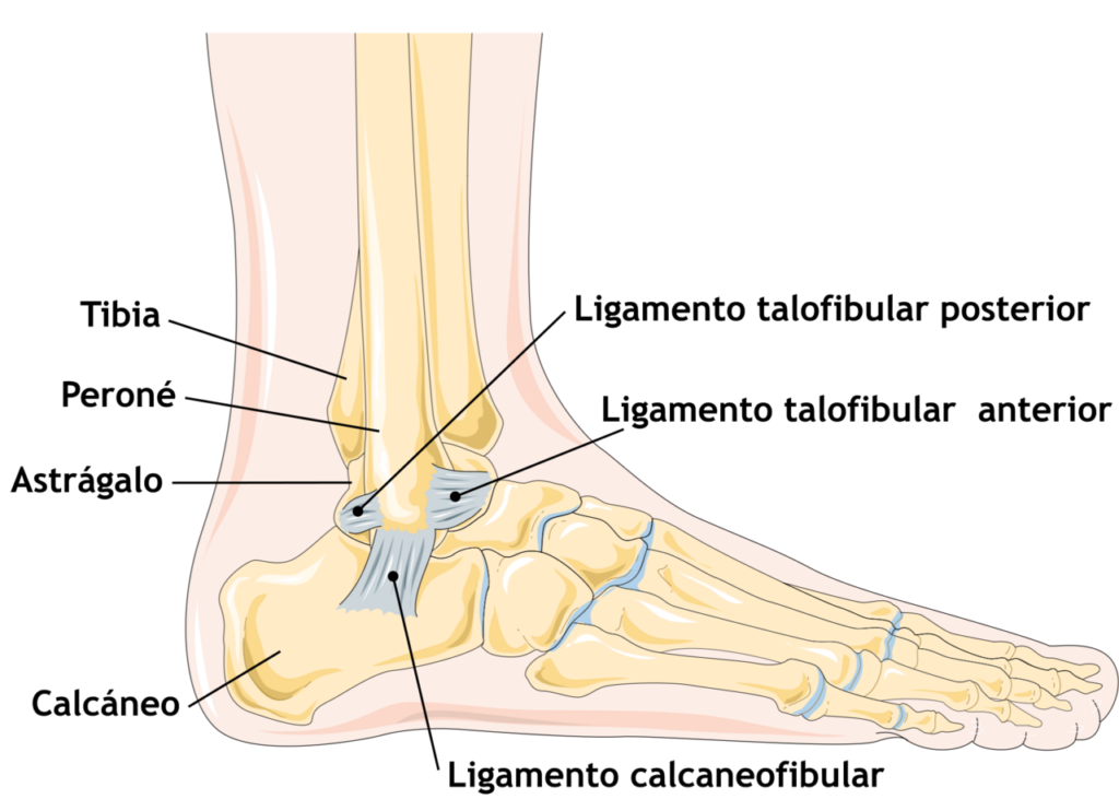
- Chronic lateral ankle instability failing rehabilitation
- Recurrent sprains with giving way despite peroneal strengthening and proprioceptive training
- Mechanical instability on anterior drawer and talar tilt tests
- Assess for hindfoot varus, peroneal tears and generalised laxity
- Stress radiographs or MRI support the diagnosis
Position
- Supine, bump under the hip, thigh tourniquet
- Image intensifier for anchor placement
Operative Steps
- Curved incision along the anterior fibula
- Protect the superficial peroneal and sural nerves
- Arthroscopy first to treat intra-articular lesions
- Divide and imbricate the ATFL and CFL, or reattach them to the fibula with anchors (Brostrom)
- Advance the inferior extensor retinaculum over the repair (Gould)
- Tendon graft reconstruction for poor tissue or failed repair
- Correct cavovarus with a calcaneal osteotomy if present
- Arthroscopic or all inside Brostrom techniques are alternatives
Postoperative Care
- Boot for about 6 weeks
- Proprioceptive and peroneal rehabilitation
- Weight bearing in the boot as comfortable
- Avoid inversion for 6 weeks
- Return to sport at 3 to 4 months
- Brace for sport in the first season
Complications
- Superficial peroneal and sural nerve injury
- Stiffness and loss of inversion
- Recurrent instability
- Wound breakdown over the lateral malleolus
- Peroneal tendon irritation
- Subtalar stiffness after nonanatomic tenodesis such as Evans or Chrisman Snook
Related Pages
Reviewed by Professor Phong Tran, Head of Orthopaedic Surgery, Western Health. Last updated 10 October 2026.