
Question
A 12 year old girl has a painful, expanding bone lesion. Her imaging is shown.
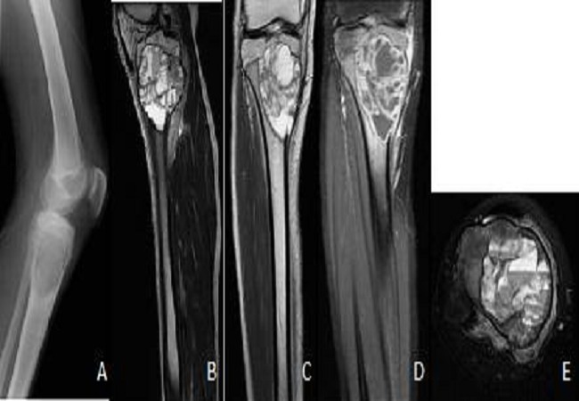
- What are the imaging features and the differential diagnosis?
- What are the pathological features?
- How is it treated?
Answer
Imaging and differential
- Eccentric, expansile, lytic metaphyseal lesion with a thin cortical shell
- Fluid levels on MRI
- Telangiectatic osteosarcoma must be excluded
- Others are simple bone cyst, giant cell tumour and chondroblastoma with secondary ABC change
- Aggressive lesions can cross the physis
Pathology
- Blood filled spaces separated by septa of fibroblasts, giant cells and reactive bone
- Primary ABC carries a USP6 gene rearrangement
- Secondary ABC arises within giant cell tumour, chondroblastoma or osteoblastoma
Treatment
- Biopsy before definitive treatment
- Intralesional curettage with a high speed burr and adjuvant, then bone graft
- Recurrence is frequent, especially in young children
- Sclerotherapy, denosumab or selective arterial embolisation for spinal and pelvic lesions
Related pages
Author Contributions
Orthofracs team
Reviewed by Professor Phong Tran, Head of Orthopaedic Surgery, Western Health. Last updated 10 October 2026.