
Question
A 30 year old woman has knee pain. Radiographs show an eccentric lytic lesion in the proximal tibial epiphysis extending to the subchondral bone, with no matrix mineralisation.
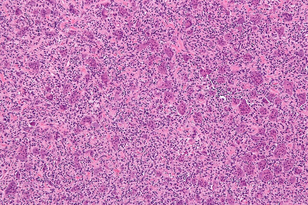
- What is the most likely diagnosis?
- How is it classified and staged?
- What are the treatment options?
Answer
Diagnosis
- Giant cell tumour of bone, a locally aggressive benign tumour
- Skeletally mature patients aged 20 to 40, around the knee in about half
- Eccentric epiphyseal lesion extending to subchondral bone with a narrow non-sclerotic margin
- Histology shows osteoclast like giant cells among mononuclear stromal cells, which carry the H3F3A mutation and express RANKL
Classification and staging
- Campanacci radiological grades 1 latent, 2 active and 3 aggressive with cortical breach
- Enneking benign staging 1 to 3
- CT of the chest, as about 2 to 5% develop benign pulmonary metastases
Treatment
- Biopsy to confirm and exclude telangiectatic osteosarcoma and brown tumour
- Extended intralesional curettage with a high speed burr and adjuvants such as phenol, liquid nitrogen or argon beam
- Fill the defect with cement (PMMA) or bone graft
- Recurrence about 10 to 20%, mostly within 2 years
- Wide resection with reconstruction for extensive destruction or recurrence
- Denosumab for unresectable tumours or to downstage, with a risk of rebound recurrence after stopping
Related pages
Author Contributions
Orthofracs team
Reviewed by Professor Phong Tran, Head of Orthopaedic Surgery, Western Health. Last updated 10 October 2026.