
Question
A 16 year old boy has a hard lump behind his knee that hurts when he kneels.
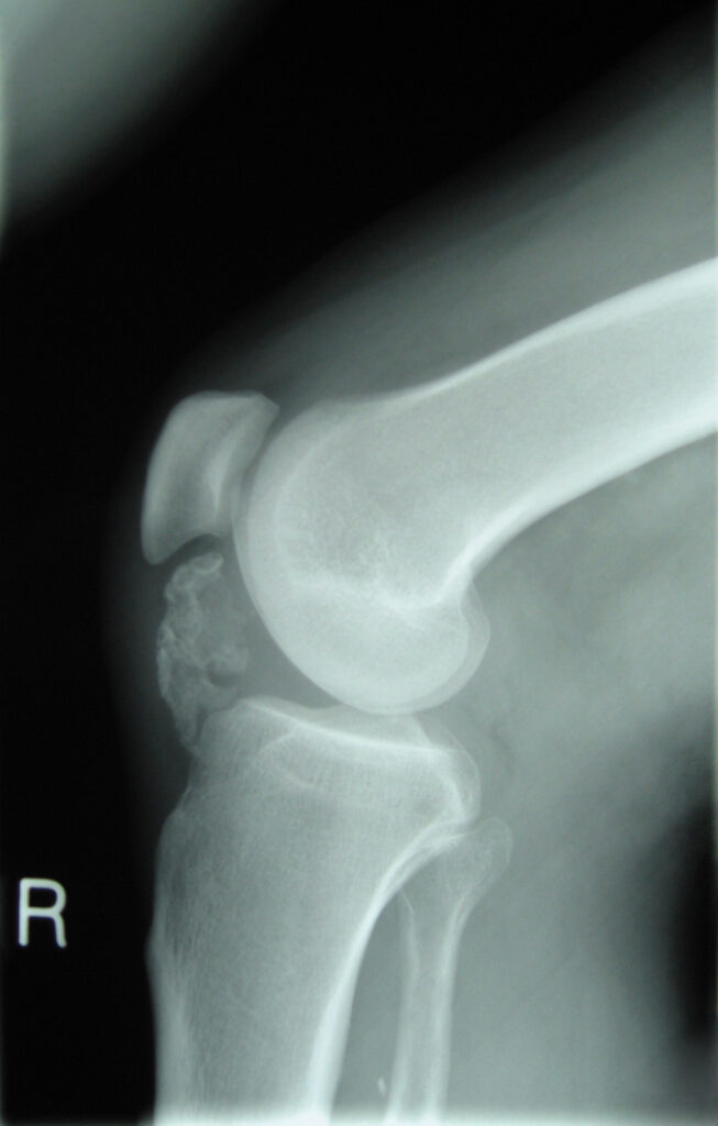
- Describe the radiograph and the pathology.
- What symptoms or signs would suggest malignant change?
- When would you excise it and how?
Answer
Radiograph
- Pedunculated bony projection from the posterior distal femoral metaphysis
- Cortex and medulla continuous with the host bone
- Points away from the joint
- Solitary osteochondroma (exostosis)
Pathology
- Most common benign bone tumour
- Displaced physeal cartilage grows at an angle to the physis
- EXT1 or EXT2 mutations in hereditary multiple exostoses
- Growth stops at skeletal maturity
Malignant change
- Under 1% for solitary lesions, higher in multiple exostoses
- New pain or growth after skeletal maturity
- Cartilage cap over 2 cm in adults on MRI
- Irregular or destroyed lesion margin, soft tissue mass
- Secondary peripheral chondrosarcoma
Excision
- Pain, mechanical symptoms, nerve or vessel compression, bursitis, cosmesis or concern for malignancy
- Excise the whole cartilage cap and perichondrium to prevent recurrence
- Delay until near maturity if possible
- Popliteal vessels and nerves at risk in this location
Related pages
Author Contributions
Orthofracs team
Reviewed by Professor Phong Tran, Head of Orthopaedic Surgery, Western Health. Last updated 10 October 2026.