
Question
An 87 year old woman has deep thigh pain at night. An incidental lesion was noted on a radiograph several years ago.
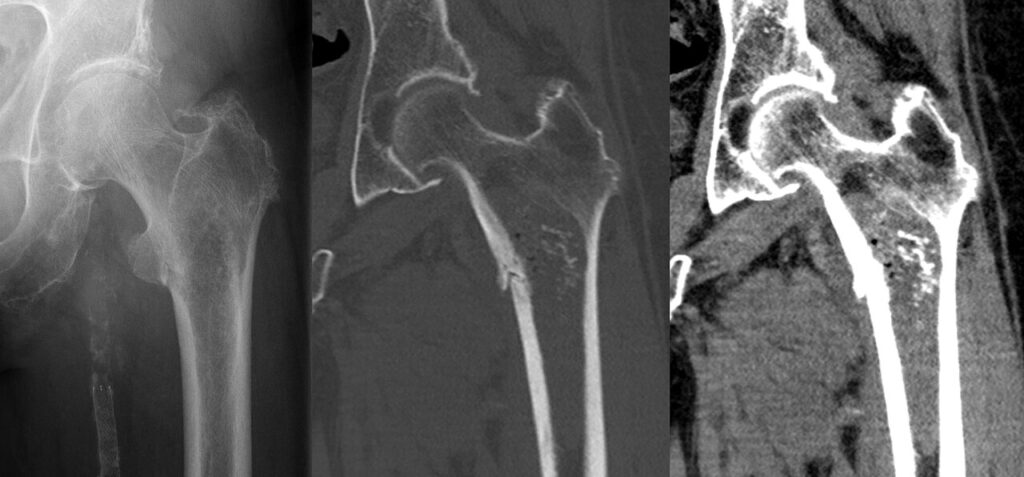
- Describe the imaging.
- What features suggest a malignant cartilage tumour?
- Outline the workup and treatment.
Answer
Imaging
- Lytic and sclerotic lesion of the proximal femur with ring and arc calcification
- Endosteal scalloping and cortical breach
- Chondrosarcoma
Features of malignancy
- Pain at rest or at night
- Endosteal scalloping over two thirds of cortical thickness
- Cortical destruction, periosteal reaction or soft tissue mass
- Proximal or axial location
- Size over 5 cm
- Growth of a known lesion
- Increased uptake on bone scan greater than the iliac crest
Workup
- Refer to a sarcoma centre before biopsy
- MRI of the whole bone
- CT chest for staging
- Core biopsy through the planned resection tract
Treatment
- Resistant to chemotherapy and radiotherapy
- Wide resection with endoprosthetic reconstruction
- Atypical cartilaginous tumours of the long bones may be treated with intralesional curettage and adjuvant
- Dedifferentiated chondrosarcoma has a poor prognosis and is treated like osteosarcoma
- Grade predicts survival
Related pages
Author Contributions
Orthofracs team
Reviewed by Professor Phong Tran, Head of Orthopaedic Surgery, Western Health. Last updated 10 October 2026.