
Definition
- A benign cystic lesion of bone that expands the cortex.
- It is the only bone lesion that derives its name from its X-ray appearance rather than its histology.
- Term first used by Jaffe & Lichtenstein in 1942
Classification
| Type | Description |
|---|---|
| Primary | Arise de novo |
| Secondary | Arise in pre-existing tumours, frequently GCTs. More than 50% of ABCs arise from preexisting lesions |
Incidence
- 2.5% of all primary bone tumours.
- Half as frequent as giant cell tumours
- M=F
- 80% in the first two decades of life
- In contrast, only 15% of patients with GCT are in the first two decades of life
Aetiology
- Unknown
- Trauma may be related
- nearly 1/2 are seen to occur in conjunction with another benign tumour and may represent a breakdown in the body’s reaction to the other tumour
Localisation
- Can involve any bone.
- 1. Proximal end of long bones
- In long bones involves the metaphysis but can cross the growth plate.
- 2. Spine
- In the spine involve the posterior elements (Differential Diagnosis here is osteoblastoma).
- 1. Proximal end of long bones
- ABC is the commonest benign tumour of the clavicle
History
- Pain
- usually mild and intermittent
- may be Rapid ↑ in pain
- Swelling
- Swelling will tend to increase until the lesion is treated
- Rarely, a pathologic fracture
- Neurological
- If the lesion is located in the vertebral column, it may cause signs and symptoms of spinal cord compression (leg weakness, bowel or bladder dysfunction).
Examination
- May be a mass or neurological symptoms if the spinal cord is compressed
Investigations
X-ray
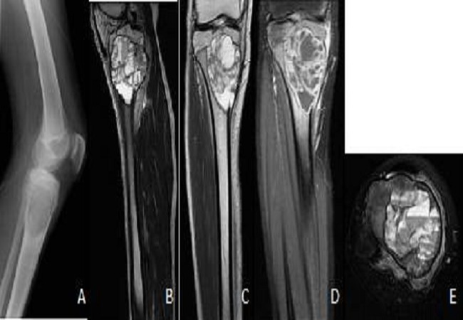
- Expansile, multilocular balloon disrupting the adjacent bone & elevating the periosteum
- Most commonly, an area of lucency situated eccentrically in the medullary cavity of a long bone.
- Less commonly, may be situated centrally within the medulla.
- Much less commonly, may arise in the cortex or periosteum.
- No matrix mineralisation is present in the lesion
- Most ABCs are completely lytic
- ABCs may cross joints & involve several bones
- particularly in the spine where several adjacent vertebrae & ribs may be involved.
- lesion tends to involve the cortex & may destroy it completely, when it may bulge out into the soft tissue where it usually forms a thin rim of calcification.
- margins can be poorly or well defined; in half of cases the X-ray appearances suggest a benign process, & in a small number of cases they may suggest malignancy.
- Periosteal new bone formation causing a buttress effect is characteristic.
- There is no matrix calcification
CT & MRI
- internal septae & fluid levels with a layering effect.
- Fluid levels (T2 weighted scans)
- Finger in balloon sign
- preservation of a cortical cuff extending for a short distance into the expanded area of destructive blowout
Pathology
Gross Pathology
- Red brown granular material (haemosiderin deposition)
- “a hole containing blood”
- blood in the lesion is not clotted
- no endothelial lining
- pressure in an ABC may be elevated to arteriolar levels
Histology
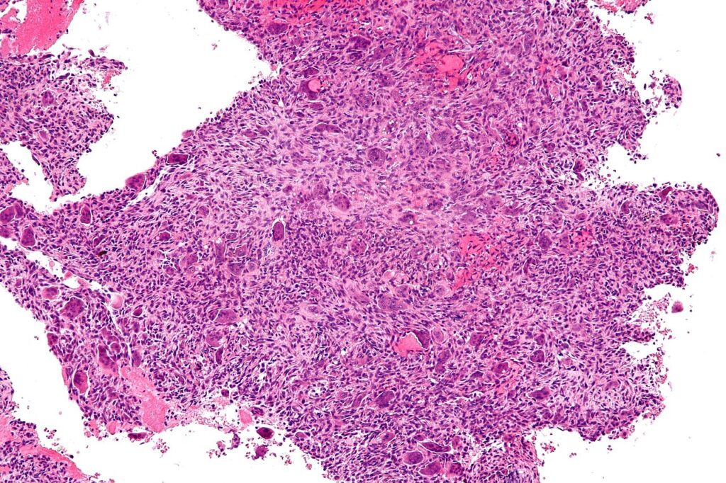
- Essential feature is cavernomatous spaces,
- with walls that lack the normal features of blood vessels, such as muscle or elastic lamina
- Thin strands of bone are often present in the fibrous tissues of the walls
- septae almost invariably contain giant cells; this helps to distinguish from unicameral bone cyst
- Solid areas contain spindle cells that are loosely arranged
- Chondroid like zones of calcification in solid portions of septae are commonly found & are relatively specific
Behaviour
- ABCs are usually aggressive lesions associated with major bone destruction, pathological fractures & local recurrence
- Spontaneous malignant transformation not recorded in the Mayo files but occurs very occasionally
Differential Diagnosis (Histology)
- Giant cell tumour
- Giant cell reparative granuloma
- Low-grade osteosarcoma
- Telangiectatic osteosarcoma – probably most difficult Differential Diagnosis
- This disease is uncommon & rarely involves the vertebrae or small bones of the hands or feet
- Renal cell carcinoma metastasis
Associated Conditions
May occur in other benign bone tumours or processes
- Giant cell tumour
- Chondroblastoma
- Osteoblastoma
- Fibrous dysplasia
- NOF
Treatment
- Intralesional curettage & bone grafting & chemical cauterization of cyst walls
- treatment of choice
- Lesions in expendable bones are excised
- Preoperative Embolisation
- If the lesion has a large soft tissue component or is large (>5-6cm) should consider preoperative embolisation
- Especially pelvis, sacrum, vertebral bodies
- Even incomplete resection may be followed by regression of the lesion
- High recurrence rate
- Recurrence tends to occur within 6-18 months. It is very rare after 2 years.
- Recurrence rate is higher in spinal tumours
- Radiotherapy
- has no role unless surgery is impossible.
- It used to be used adjuvantly but no longer is used to minimise the risk of sarcomatous transformation
Prognosis
- 95% of patients cured
- ↑ rate of recurrence in younger patients
- Children with open physes are much more prone to local recurrence (up to 50%)
Reviewed by Professor Phong Tran, Head of Orthopaedic Surgery, Western Health. Last updated 10 October 2026.