
Question
A 25 year old woman has a lesion found on a radiograph taken after an ankle sprain.
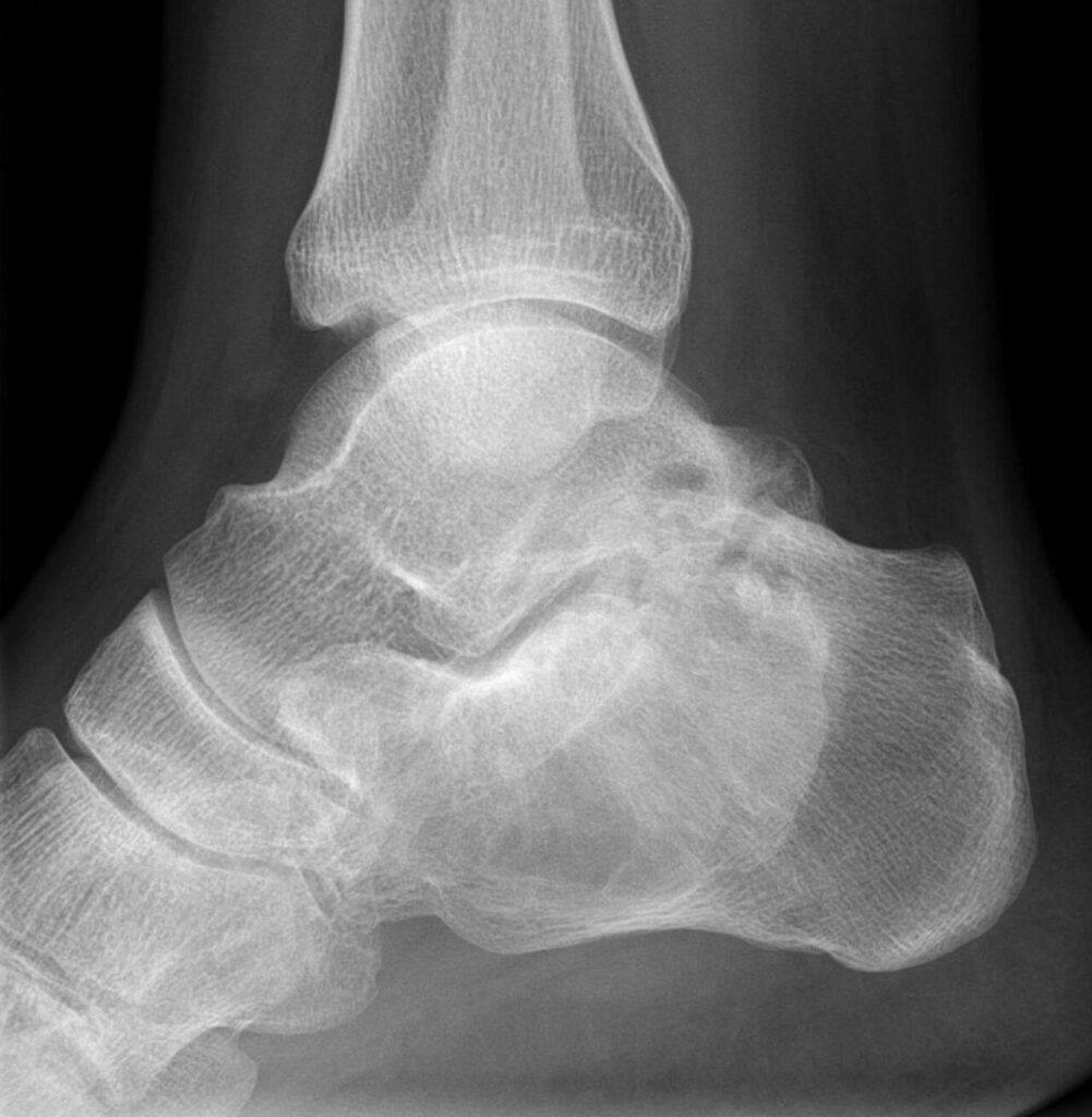
- What is the likely diagnosis and its features?
- What syndromes are associated?
- How is it managed?
Answer
Diagnosis
- Fibrous dysplasia
- Well defined lesion with a ground glass matrix
- Proximal femoral involvement can cause a shepherd’s crook deformity
- Activating somatic GNAS mutation
- Histology shows woven bone trabeculae (Chinese letters) without osteoblastic rimming
- Monostotic in most patients, often in the proximal femur, ribs or skull
Associated syndromes
- McCune-Albright syndrome, with polyostotic disease, irregular cafe au lait spots and precocious puberty
- Mazabraud syndrome, with intramuscular myxomas
Management
- Observation for asymptomatic lesions
- Bisphosphonates for bone pain
- Fracture or deformity treated with intramedullary fixation and cortical allograft, as cancellous graft resorbs
- Malignant change is rare
- Bone scan shows the extent of polyostotic disease
Related pages
Author Contributions
Orthofracs team
Reviewed by Professor Phong Tran, Head of Orthopaedic Surgery, Western Health. Last updated 10 October 2026.