
Anatomy
- Branch of the common peroneal nerve at the fibular neck
- Runs in the lateral compartment between the peronei and EDL
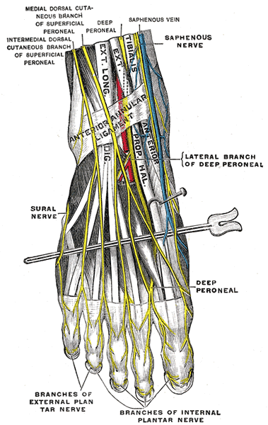
- Pierces the deep fascia about 10 to 12 cm above the lateral malleolus
- Divides into medial and intermediate dorsal cutaneous nerves
- Course varies and in some people it runs in the anterior compartment
Motor Supply
- Peroneus longus
- Peroneus brevis
Sensory Supply
- Distal anterolateral leg
- Dorsum of the foot except the first web space
Clinical Features
- Entrapment where it exits the fascia causes dorsal foot paraesthesia worse with activity
- Associated with fascial defects, muscle herniation and ankle sprains
- Plantarflexion with inversion makes the nerve visible under the skin
- Tinel sign at the fascial exit point
Investigations
- Nerve conduction studies when the diagnosis is unclear
Differential Diagnosis
- L5 radiculopathy
- Common peroneal nerve compression at the fibular neck
- Exertional lateral compartment syndrome
Management
- Fascial release for entrapment
- Neuroma excision and burial
- Mark the nerve on the skin before anterolateral portal placement
- Neurolysis works best when a fascial band is found
Structures at Risk
- Anterolateral ankle arthroscopy portal, the commonest nerve injury in ankle arthroscopy
- Proximal end of lateral fibular plates
- Lateral compartment fasciotomy
- Lateral approach to the distal fibula, where the nerve crosses obliquely
Reviewed by Professor Phong Tran, Head of Orthopaedic Surgery, Western Health. Last updated 10 October 2026.