
Investigations
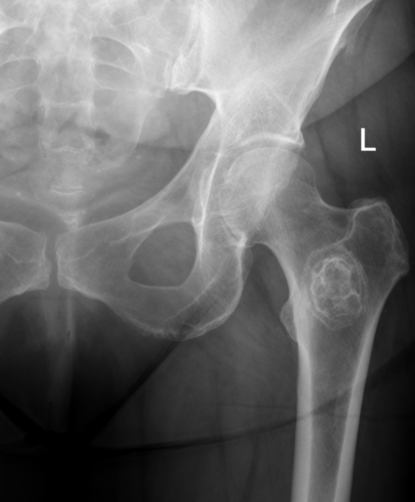
- Orthogonal radiographs of the whole bone are the most useful first test
- Aim to class a lesion as aggressive or non-aggressive with a short differential
- Narrow zone of transition indicates slow growth, wide indicates rapid growth
- Chondroid matrix shows rings and arcs or popcorn calcification
- Osteoid matrix is cloud-like or ivory, as in osteosarcoma
- Fibrous matrix shows ground glass, as in fibrous dysplasia
- Absent matrix does not exclude malignancy
- Solid smooth periosteal reaction indicates a slow process
- Lamellated onion skin reaction in Ewing sarcoma and infection
- Sunburst or hair on end spicules indicate aggressive growth
- Codman triangle indicates aggression but is not specific to malignancy
- Endosteal scalloping over two thirds of cortex favours chondrosarcoma over enchondroma
- Soft tissue mass indicates an aggressive process
Classification
Lodwick
| Grade | Description |
|---|---|
| IA | Geographic with sclerotic rim |
| IB | Geographic, well defined, no sclerotic rim |
| IC | Geographic with ill defined margin |
| II | Moth-eaten |
| III | Permeative |
Original publication Lodwick GS, Wilson AJ, Farrell C, Virtama P, Dittrich F. Determining growth rates of focal lesions of bone from radiographs. Radiology. 1980;134(3):577-83.
- IA and IB are non-aggressive
- Moth-eaten and permeative suggest Ewing sarcoma, lymphoma, osteosarcoma, myeloma or osteomyelitis
Differential Diagnosis
- Under 5 years, neuroblastoma metastasis, LCH and infection
- 5 to 20 years, osteosarcoma, Ewing sarcoma, bone cysts, NOF, chondroblastoma and osteochondroma
- 20 to 40 years, giant cell tumour of bone
- Over 40 years, metastasis, myeloma, lymphoma and chondrosarcoma
- Multiple lesions, metastasis, myeloma, lymphoma, LCH, multiple enchondromas and HME
- Multiple lesions also in fibrous dysplasia, vascular tumours and infection
- Infection, LCH and ABC can look aggressive
- Combine imaging with age, history and histology
Reviewed by Professor Phong Tran, Head of Orthopaedic Surgery, Western Health. Last updated 10 October 2026.