
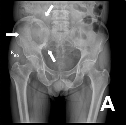
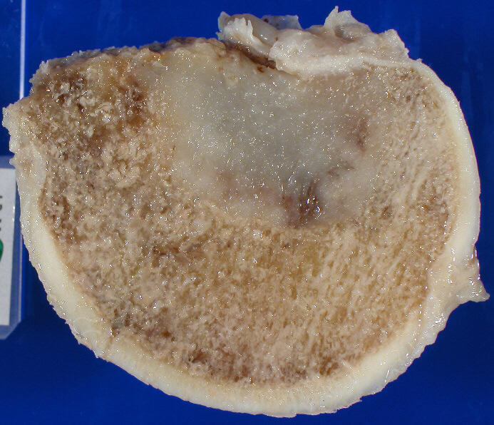
Bony Metastases
Survival
Median survival after diagnosis of bony mets:
- Thyroid: 48 mth
- Prostate: 40 mth
- Breast: 24 mth
- Myeloma: 20 mth
- Renal: < 6 mth. If solitary met, there is a chance of long term survival
- Lung: < 6 mth
Radiotherapy
- Breast & prostate more responsive than lung
Indications for Prophylactic Fixation
- Considerations:
- Pain
- Persistent pain despite radiation
- Location of lesion
- Involvement of subtrochanteric region ↑ risk
- Any lesion between lesser trochanter & femoral head causing functional pain or > 2.5 cm should be fixed
- Avulsion of lesser trochanter
- Lesion size
- Many descriptors used
- Lesion > 2.5 cm involving femoral cortex
- > 50% femoral cortical involvement (100% incidence of pathologic fracture)
- Many descriptors used
- Pain
- Mirels system
- Pain
- Mild (1)
- Moderate (2)
- Severe (3)
- Location of lesion
- Upper limb (1)
- Lower limb (2)
- Peritrochanter (3)
- Size of lesion
- < 1/3 (1)
- 1/3-2/3 (2)
- > 2/3 (3)
- Type of lesion
- Blastic (1)
- Mixed (2)
- Lytic (3)
- Assessment – score out of 12:
- < 7, not at risk of fracture
- 8, 15% risk of fracture
- 9, 33% risk
- >=9, should be used as an indication for fixation
- Pain
Adjuvant Therapy for Metastatic Bone Disease by Dr Vikram David
Management of Metastatic Bone Disease
Reviewed by Professor Phong Tran, Head of Orthopaedic Surgery, Western Health. Last updated 10 October 2026.