
Epidemiology
- Children
- Most common, high grade, central, affects children
- 85% of osteosarcomas
- M:F – 1.5:1
- Location
- 90% in ends of long bones
- 50% around knee
- distal femur 34%
- proximal tibia 17%
- femoral shaft 5%
- proximal femur 5%
- proximal humerus 9%
Clinical Features
- Localised swelling & pain in children (usually teens)
- Pain & mass
- Warmth
- No systemic symptoms
- Mean symptom duration is 4/12
- 10% metastases at presentation
- Pathological fracture occasionally
Pathology
- Dependent on predominant type of matrix produced
- Lichenstein’s classification
- Conventional
- Bony (50% of all central OS)
- Cartilaginous
- Fibrous
- Telangiectactic
- Small cell
- Giant cell (fibrohistiocytic)
- Conventional
- Therefore gross & micro appearance varies
- Osteosarcoma is the great histological imitator among bone tumours just as Ewings sarcoma is the great radiological imitator among bone tumours
Gross
- Starts with intramedullary focus
- Bony with areas of focus & haemorrhage
- Skip lesions common 5-20%
- Grows up & down medulla
- Penetration of cortex common
- Penetration of epiphysis less common
- Involvement of joint rare
Histology
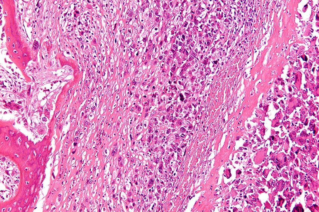
- Diagnosis depends on production of malignant osteoid by the tumour
- Cellular areas with microscopic trabeculae of bone between cells
- Pleomorphic spindle cells with
- Hyperchromatic nuclei
- Atypical mitotic figures
- May also see anaplastic cell matrix & mix of fibrous, cartilaginous & bony tissues
- Conventional form
- Osteoblastic
- Prominent osteoid
- Chondroblastic
- Prominent cartilage
- Fibroblastic
- Prominent fibrous tissue
- Looks like fibrosarcoma
- Osteoblastic
- Telangiectatic form (< 5%)
- Similar to ABC
- Large lytic defect filled with blood filled cavity
- Dilated vascular channels with multinucleated giant cells
- Anaplastic sarcomatous stroma with bone formation
- Worse prognosis than conventional form
- Small cell form
- Similar to Ewing’s sarcoma
- Nests of small cells with spindle arrangement
- Sparse osteoid
- Worse prognosis than conventional form
- Responds to chemotherapy like PNET
- Fibrohistiocytic variant (giant cell variant)
- Similar to MFH
- Higher age group in 3rd decade
- Pleomorphic spindle cells & multinucleated giant cells
- Osteoid with inflammatory matrix
Central Osteosarcoma Low Grade
- Unusual
- Older patient
- M=F
- Bone around the knee most common sites
- Spindle cells with appearance of fibrous dysplasia & desmoid
- Radiology more ominous than the histology
Metastatic Spread
- Primarily haematogenous
- Lung mets can be treated by chemotherapy & surgical excision
- Bone mets have very poor prognosis
- Up to 80% have at least micrometastatic spread on presentation
- Multifocal OS very rare (? actually represents metastatic OS)
Investigations
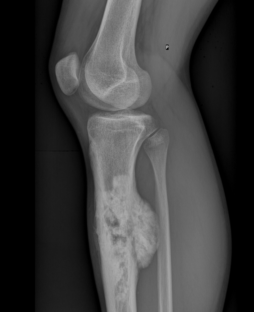
X-ray
- Definitive Diagnosis in 55%
- Differential Diagnosis in 35%
- Wilner’s classification
- Sclerotic 30%
- Lytic 20%
- Mixed 45%
- Central & destructive lesion in metaphysis
- Involves medullary canal
- Lytic & Blastic
- Permeative cortical destruction
- Periosteal reaction of rapid growth
- Codman’s Triangle (benign reactive bone which should be avoided in biopsy)
- Sunburst Spicules
- Soft tissue component
- Fluffy neoplastic bone
- Rarely in diaphysis or even epiphysis
- Soft tissue extension the rule
CT Scan
- Defines extraosseous component
- Identifies neoplastic bone in soft tissues
- CT lung
- 10% of patients have pulmonary mets on presentation
- Best resolution for metastases
MRI
- Best for
- Intraosseous limit
- Soft tissue component
- Relationship to NV structures
- Skip lesions (metastasis outside reactive zone)
- Whether joint is involved (rare)
Bone Scan
- Identifies
- Skip lesions
- Metastases
- Polyostotic disease
- Pulmonary metastases
Laboratory
- ALP & LDH can be ↑
- Worst prognosis
- May be mild ↑ in ESR
Differential Diagnosis
- Bone island
- Osteoma
- Osteoblastoma
- Ewing’s sarcoma
- Chondrosarcoma
- Fibrosarcoma
- Myositis ossificans
- Osteochondroma
- Juxtacortical chondroma
- Fracture callus
Treatment
Chemotherapy
- Systemic treatment
- Goals of chemotherapy
- Shrink tumour size
- Treat micrometastases
- Assess tissue response
- Allow Limb Salvage Surgery (LSS)
- Time to manufacture custom prosthesis
- Neoadjuvant & Adjuvant (pre & post-op chemotherapy)
- Marked improvement in the 5 year survival rates with this
- Response to chemotherapy best indicator of survival
- Necrosis Response (Huvos Grade: Grade 1 < 50%, Grade IV > 90%)
- > 90% necrosis correlates with good prognosis
- Greater the necrosis » better the survival
- Some osteosarcomas have P-Glycoprotein Pump
- Which removes chemotherapy from the cell
- 2 cycles Preoperative
- Rosen T 10 Regime outdated
- Current regimen “MACI”
- MTX
- Adriamycin
- Cisplatin
- I-Phosphamide
- Postoperative regimen
- Similar
- Can add VP16 & CPM (? Only if surgical excision shows > 10% tumour viability)
Surgery
- Usually 3/12 after diagnosis
- Usually 2/52 after end neoadjuvant chemotherapy
- Consists of wide excision
- 7cm proximally, 5cm distally
- Can be in form of amputation or limb salvage procedures
- Reconstructive Technique
- Endoprosthesis ~ Best Results
- Arthrodesis
- Allograft
- DXRT to prevent local recurrence
- Adjuvant Chemotherapy for systemic treatment
- Reasons for amputation
- Soft-tissue & neurovascular involvement
- Gross leg-length inequality (young child)
- Infected tumour
- Unstable pathological fracture
- Disadvantaged situation (environment, patient variables)
- Poor outcome with any local recurrence after surgery
- Poor outcome in presence of pathological fracture
- Isolated Lung metastasis can be resected with good long term outcome
Outcome
- Prior to 1971
- 20% 5 year survival rate
- Now
- 70% 5 year survival rate (80% some centres)
- Due to
- Improved resection techniques
- Adjuvant chemotherapy (Mayo clinic – 40% 5 year survival without chemo)
- Resection of lung metastases early (some thoracic surgeons happy to do multiple metastectomies for osteosarcoma)
- Limb salvage now viable option
- Local recurrence rate the same in distal femoral OS with limb salvage vs AKA
- 74% LSS vs 64% amputation at distal femur
- Local recurrence rates are the same at 5-10%
- Local recurrence rate the same in distal femoral OS with limb salvage vs AKA
- Local Recurrence
- Incidence 5-10%
- Survival 5-10% at 5 years
- Treat with local measures only
- Dickinson says » recurrence = death
- Worse prognosis if
- > 15cm (3x)
- Symptoms < 6/12
- Age < 10 years
- Males
- Elevated ALP & LDH
- Osteoblastic/ Chondroblastic
- Proximal or Central lesion
- Pathological fracture
- NV involvement
- Late Presenter
- Paradoxically better survivor
- Less aggressive tumour
- Main indicator of survival is nature of tumour
- Rather than nature of treatment
- Death due to
- Local recurrence
- Lung secondaries
Reviewed by Professor Phong Tran, Head of Orthopaedic Surgery, Western Health. Last updated 10 October 2026.