
Investigations
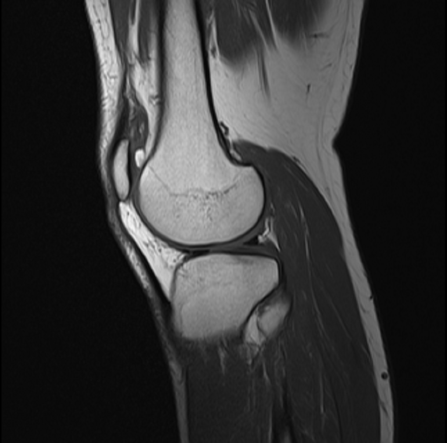
- MRI is the investigation of choice for local staging
- Radiographs are the best first test to characterise a bone lesion
- CT is superior for matrix, cortex and fine periosteal reaction
- Perform MRI before biopsy, as haemorrhage and oedema blur margins
- T1 best shows marrow replacement and true extent for resection length
- Fat suppressed T2 or STIR overestimate extent by including oedema
- Contrast T1 fat suppressed separates viable tumour from necrosis and targets biopsy
- Gradient echo shows haemosiderin blooming, as in tenosynovial giant cell tumour
- Diffusion and dynamic contrast imaging assess chemotherapy response and recurrence
- Image the whole bone with a long coronal T1 to detect skip metastases
- Skip lesions occur in osteosarcoma and change the resection extent
- Image the whole compartment for soft tissue tumours
- Calcification and matrix are poorly shown
- Metal artefact complicates surveillance, helped by metal reduction sequences
Anatomy
- Longitudinal marrow extent and distance from the joint surface
- Physeal and epiphyseal involvement decides joint sparing resection in children
- Cortical breach and soft tissue mass size
- Intra-articular extension through cartilage, capsule or cruciate attachments
- Preserved fat plane suggests a free vessel
- Encasement beyond about 180 degrees suggests vessel involvement
- Compartmental status for Enneking staging
Differential Diagnosis
- Most tumours are low T1 and high T2
- High T1 in lipoma, well differentiated liposarcoma, haemangioma, subacute haemorrhage and melanin
- Low T2 in desmoid, haemosiderin and dense mineralisation
- Fluid-fluid levels suggest ABC but also occur in telangiectatic osteosarcoma
- Cartilage tumours are lobulated, bright on T2, with ring and arc enhancement
- Peritumoural oedema can mimic tumour extension
- Signal characteristics are rarely diagnostic alone
Management
- MRI assesses neoadjuvant response
- MRI surveys the tumour bed for local recurrence after resection
Reviewed by Professor Phong Tran, Head of Orthopaedic Surgery, Western Health. Last updated 10 October 2026.