
Definition
- Disease characterised by the replacement of normal bone & marrow by fibrous tissue & irregular minute bone spicules
- Fibrous dysplasia, precocious puberty & café-au-lait patches form the McCune-Albright syndrome
Epidemiology
- Patients tend to be in the second or third decade.
- More common in females
- Polyostotic fibrous dysplasia is much more common in females
Site
- Most common site is the proximal femur
- ribs & jaw are commonly involved. Involvement of the jaw may result in leontiasis & cherubism
- lesion is typically intramedullary & diaphyseal
Aetiology
- Failure of skeletal maturation, not hereditary
Clinical Features
- Many patients are asymptomatic
- Patients may develop deformities,
- particularly with polyostotic fibrous dysplasia
- such as the classic shepherd’s crook deformity of the proximal femur
- Fractures develop on the tension side of affected bones
- Associations
- Hyperthyroidism
- hyperparathyroidism
- hypophosphataemic rickets
- osteomalacia
Investigations
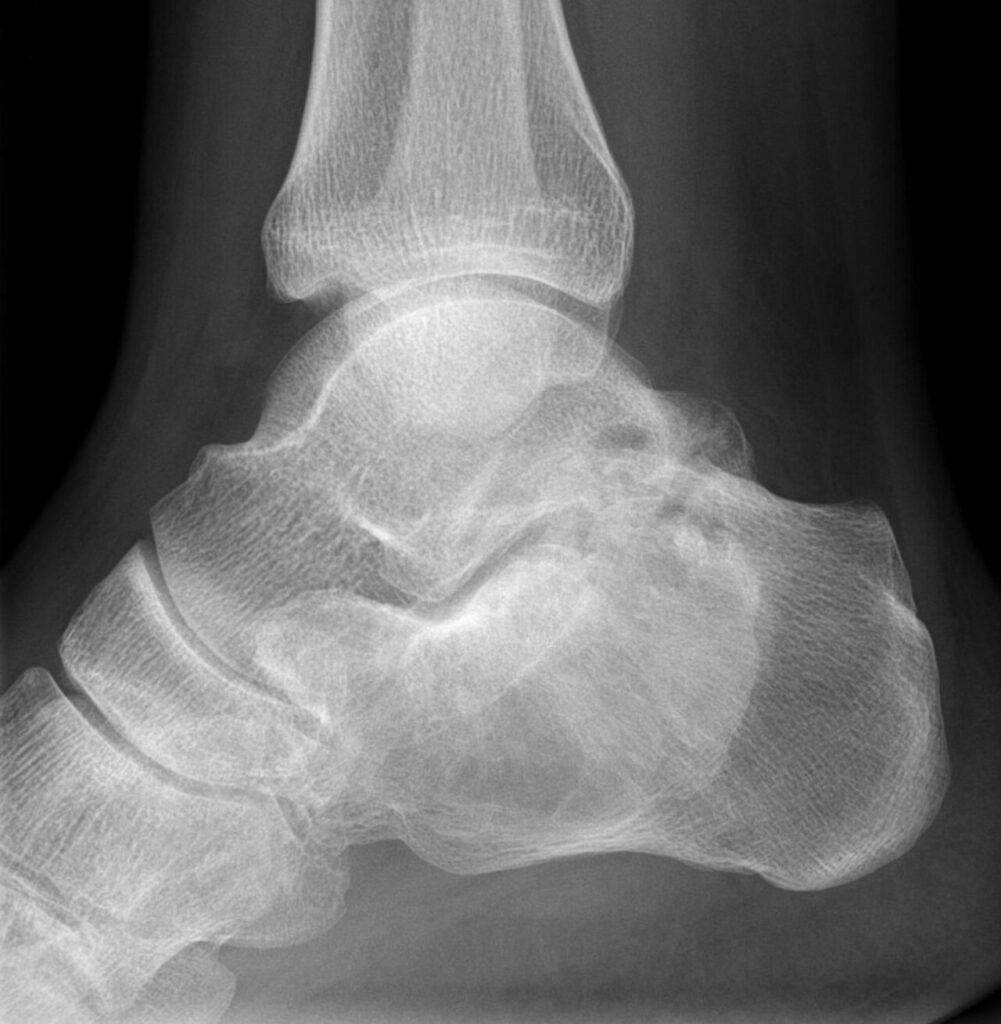
X-ray
- Well demarcated zone of rarefaction, often surrounded by a rim of sclerotic bone
- Expansion with thinning of the cortices is particularly likely in thin bones such as the ribs
- There may be a hazy appearance, classically called a ground glass appearance
MRI
- Low T1, variable T2. Cartilage is sometimes seen which will lead to ↑ T2
Bone Scan
- may be ↑ or ↓
Laboratory Findings
- Increased alkaline phosphatase
Pathology
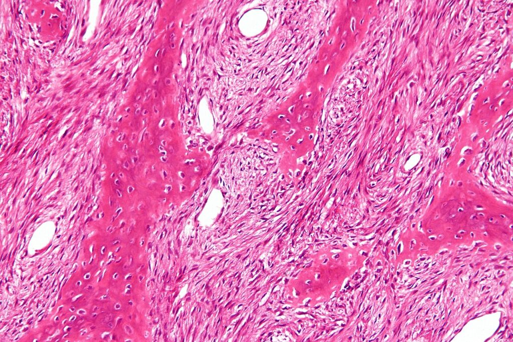
- Composed of dense fibrous tissue, surrounding bony trabeculae arranged in a meaningless fashion, known as Chinese alphabet.
- Collections of foam cells forming little islands amongst the fibrous tissue are almost always seen
- Giant cells may also be seen
Treatment
- Usually expectant.
- lesions tend to stop growing at skeletal maturity
- however reactivation may occur during pregnancy or during oestrogen therapy
- Surgery
- Large lesions in weight bearing bones need curettage & grafting, often with internal fixation
- use of cortical allograft should be considered, because it provides structural support & is slower to resorb
- Sarcomas may arise from fibrous dysplasia
Reviewed by Professor Phong Tran, Head of Orthopaedic Surgery, Western Health. Last updated 10 October 2026.