
Principles
- Imaging used for four aspects
- Make the diagnosis
- Stage the lesion
- Plan the treatment
- Staging
- Biopsy
- Assess the response
Enneking’s Questions
- What is the location of lesion?
- What is the lesion doing to the bone?
- What is the bone’s reaction to the lesion?
- What is the tumour matrix?
Imaging Modalities
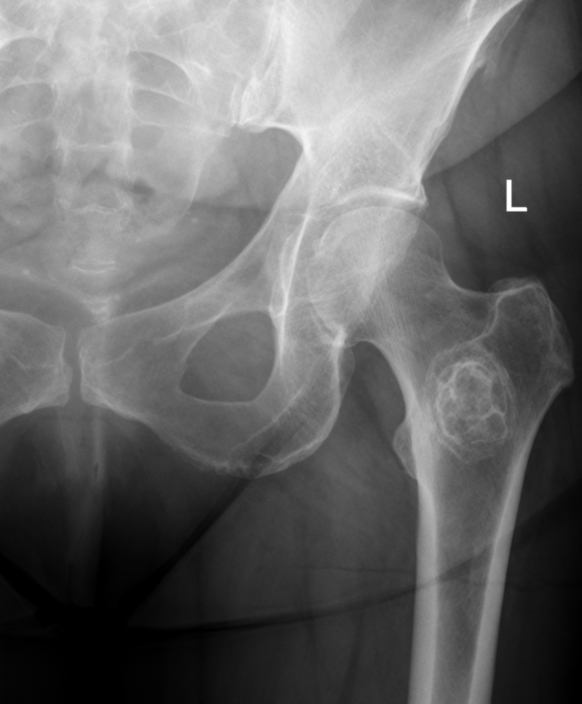
X-ray
- Initial examination
- Main-stay
- Evaluation of
- Location
- Margin
- Periosteal Reaction
- Matrix
- Evaluation of
Bone Scan
- Most lesions have ↑ uptake
- Screening tool
- Non-anatomical
- False negative with
- Inactive benign tumours
- Myeloma
- Eosinophilic Granuloma
- Haemangioma
- Melanoma
- Crude indicator of extent of lesion & response to treatment
- Main role is demonstration of multiple lesions
CT Scan
- Best for assessing mineralisation & bony details
- Benign bone tumours
- Violation of cortex
- Matrix mineralisation
- Shows local extent of tumour
- Intraosseous
- Extension into soft tissue
- Shows areas that plain XR visualise poorly
- Spine
- Pelvis
MRI
- Best for assessing soft tissue
- Very sensitive for
- Soft tissue tumours
- Soft tissue extension
- Marrow involvement
- Joint & Epiphyseal involvement
- Shows relationship to neurovascular bundle well
- May be oversensitive
- Oedema (reactive zone) vs Tumour
Diagnosis
- Mainstay is plain film
- Other modalities contribute
- CT Scan
- Defines architecture of lesion
- MRI Scan
- Determines extent of disease (can be over-sensitive)
- Bone Scan
- Locates other lesions
- CT Scan
Spread
- MRI is best
- Intramedullary spread
- T1 sequence
- T1 with Fat Suppression/ Inversion Recovery
- T1 sequence
- Cortical breakthrough
- T2 sequence
- Soft tissue spread
- T2 sequence
- Relationship to vessels
- T2 sequence
- Intramedullary spread
Metastases
- Lungs
- CXR – see mets >/= 1cm
- CT Scan – see mets >/= 2mm
- Bone
- Bone scan
Radiological Diagnosis of Bone Tumours
Watt Criteria (1985)
| Question | Interpretation |
|---|---|
| 1. Solitary or multiple? | Multiple lesions more likely to represent metastatic lesions or systemic disorder. Solitary more often primary tumour |
| 2. What type of bone involved? | Metastatic disease presents most in axial skeleton. Osteoid osteoma rarely in intramembranous bones |
| 3. Where is lesion in bone? | Fibrous cortical defect is cortical in location. Chondroblastoma epiphyseal. GCT is subarticular in mature skeleton |
| 4. Are margins well or ill defined? | Slow growing tumour has short zone of transition with normal bone. Aggressive tumours more permeative |
| 5. Is there a bony reaction? | More indolent the greater the sclerosis |
| 6. Does lesion contain calcification? | Useful signs of cartilage tumour |
Reviewed by Professor Phong Tran, Head of Orthopaedic Surgery, Western Health. Last updated 10 October 2026.