
Definition
- Bowed legs, with knees apart when the ankles touch
- Most cases in young children are physiological and resolve spontaneously
Anatomy
- Salenius and Tunnessen described normal tibiofemoral angle development
- Varus at birth
- Neutral by 18 to 24 months
- Physiological valgus peaks at 3 to 4 years
- Adult mild valgus by about 7 years
Aetiology
- Infantile and adolescent Blount disease
- Nutritional or X-linked hypophosphataemic rickets
- Skeletal dysplasias such as achondroplasia and metaphyseal chondrodysplasia
- Post-traumatic or post-infective physeal arrest
- Focal fibrocartilaginous dysplasia
Clinical Features
- Persistence or worsening beyond 2 years suggests pathology
- Asymmetry, severe deformity, short stature or dysmorphism
- Lateral thrust of the knee in gait
- Family history or dietary risk for rickets
Classification
- Blount disease affects the posteromedial proximal tibial physis, causing varus, procurvatum and internal tibial torsion
- Infantile type before 4 years, usually bilateral, linked to early walking, obesity and African descent
- Adolescent type after about 10 years, more often unilateral, in obese children, may include distal femoral varus
- Langenskiold six stages, from medial metaphyseal beaking (I) to a medial physeal bony bridge (VI)
Investigations
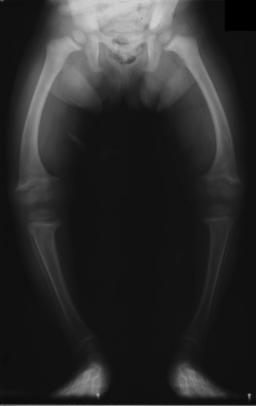
- Standing long leg radiograph when pathology is suspected
- Drennan metaphyseal-diaphyseal angle, between the metaphyseal beak line and a perpendicular to the tibial shaft
- Less than 11° likely physiological
- Greater than 16° strongly associated with Blount disease
- Rickets bloods are calcium, phosphate, ALP, vitamin D and PTH
- Rickets radiographs show widened, cupped and frayed physes
Management
- Physiological bowing, reassurance and observation
- Rickets, medical treatment first, surgery for residual deformity after metabolic control
- Infantile Blount stages I to II under 3 years, KAFO
- Progressive disease, proximal tibial valgus osteotomy with slight overcorrection, ideally before 4 years
- Later stages may need bar resection, medial plateau elevation and osteotomy
- Lateral hemiepiphysiodesis for milder disease with growth remaining
- Adolescent Blount, guided growth or osteotomy, often gradual correction in a circular frame
Reviewed by Professor Phong Tran, Head of Orthopaedic Surgery, Western Health. Last updated 10 October 2026.