
Definition
- Patella with an accessory ossification centre that fails to fuse to the main body
- Fragment joined by a fibrocartilaginous synchondrosis
Anatomy
- Patella ossifies from about 4 to 6 years
- Accessory centres appear later in childhood and normally fuse
Classification
Saupe
| Type | Location |
|---|---|
| I | Inferior pole |
| II | Lateral margin |
| III | Superolateral pole, the most common |
- Vastus lateralis attaches to the superolateral fragment
Epidemiology
- More common in boys
- Bilateral in about half
Clinical Features
- Usually incidental and asymptomatic
- Symptoms in adolescent athletes, often with lateral patellar pain
- Pain may follow injury or overuse disrupting the synchondrosis
- Tenderness over the fragment and synchondrosis
Investigations
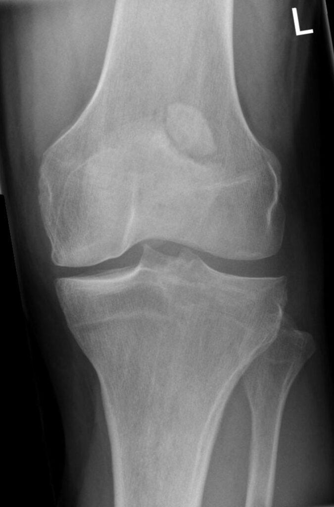
- AP and skyline radiographs show a fragment with smooth sclerotic margins
- Contralateral radiographs often show the same pattern
- MRI shows oedema across a symptomatic synchondrosis
Differential Diagnosis
- Acute patellar fracture with sharp irregular edges
- Patellar sleeve fracture
- Sinding-Larsen-Johansson disease
- Dorsal defect of the patella
Management
- Asymptomatic, no treatment
- Symptomatic, activity modification and NSAIDs
- Injury to the synchondrosis, immobilisation in extension for several weeks
- Persistent pain, excision of a small fragment
- Large fragments may need fixation
- Vastus lateralis release is an alternative for superolateral fragments
- Surgery rarely needed
Reviewed by Professor Phong Tran, Head of Orthopaedic Surgery, Western Health. Last updated 10 October 2026.