
Definition
- Knock knee with the knees touching and the ankles apart
- Intermalleolar distance over about 8 cm after age 7 is abnormal
Pathology
- Physiological alignment is varus at birth, neutral by about 2 years and maximal valgus at 3 to 4 years
- Adult alignment reached by about 7 years
Aetiology
- Rickets and renal osteodystrophy
- Skeletal dysplasias such as Morquio syndrome and multiple epiphyseal dysplasia
- Post-traumatic valgus after a proximal tibial metaphyseal fracture (Cozen)
- Physeal injury, osteochondroma and neurofibromatosis
Clinical Features
- Pathological if unilateral, progressive, severe, associated with short stature, or persisting after 7 years
- Measure intermalleolar distance with knees together
- Check for rotational deformity and patellar instability
Investigations
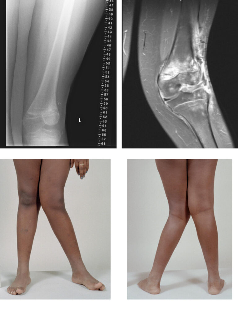
- Standing long leg X-ray to measure the mechanical axis and locate the deformity
- Calcium, phosphate, alkaline phosphatase and vitamin D if rickets is suspected
- Locate the deformity in the femur or tibia before planning surgery
Management
- Physiological valgus needs reassurance only
- Guided growth with medial tension band plates in the distal femur or proximal tibia while growth remains
- Corrective osteotomy after skeletal maturity
- See Blount’s Disease for the varus counterpart
- Physiological valgus needs reassurance only
- Treat rickets medically, which often corrects mild deformity
- Corrective osteotomy after skeletal maturity, usually distal femoral varus osteotomy
- Guided growth with a tension band plate for persistent pathological valgus in growing children
Reviewed by Professor Phong Tran, Head of Orthopaedic Surgery, Western Health. Last updated 10 October 2026.