
Definition
- Joint inflammation due to infection by micro-organisms
Incidence
- All age groups
- Usually children
- 50% < age 3
- M=F
- Any joint
- Knee is most common joint affected overall
- Infants
- Hip
- Children
- Knee
- Adults
- Large Joints
- IVDU
- SCJ & SIJ
- Infants
Pathogenesis
- Two Routes
- Haematogenous
- Distant focus
- Seeds synovial membrane
- Direct
- Extension
- Osteomyelitis
- Neonates (70% have OM & septic joint) & children
- From adjacent focus of OM
- Intra-articular metaphysis
- Proximal femur, humerus, elbow
- Osteomyelitis
- Extension
- Transphyseal vessels in neonates
- Overlying Soft tissue Infection
- Inoculation
- Penetrating injury
- Iatrogenic
- Haematogenous
Predisposition
- Three factors
- Host
- Immunodeficiency from diabetes, alcohol, malnutrition, etc
- Site
- Previous joint trauma, RA, previous HCLA, etc
- Bacteria
- Host
Aetiology
- Bugs vary with age
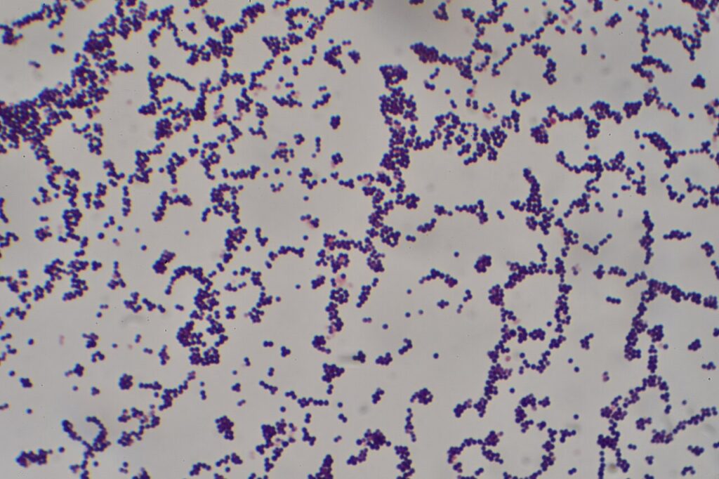
- Staphylococcus aureus most common
- Unknown in 1/3
- Community vs Hospital-acquired
- Neonates
- Hospital-acquired 60%
- E coli & Candida
- S aureus
- Group B Streptococcus
- Typically premature or unwell
- Community-acquired
- S aureus
- Group B Streptococcus
- Usually well
- 2/3 Osteomyelitis
- Hospital-acquired 60%
- Infants & Children < 3 years
- Staphylococcus aureus > Haemophilus Influenzae
- Streptococcus pyogenes
- Streptococcus pneumoniae
- E coli
- Proteus
- Pseudomonas
- Children >3 years
- Like adults
- Adults
- Staphylococcus aureus > Streptococcus (pyogenes & pneumoniae) > Gram negative (E coli, Proteus, Klebsiella, Pseudomonas)
- IVDU – Gram negative
- Sexually active – Neisseria gonorrhoeae
- Prosthetic joint – Staph epidermidis
Pathology
- Synovium oedematous & hyperaemic
- Increased cloudy synovial fluid
- 2/7
- Frank pus
- Cartilage destruction
- Starts at areas of joint contact
- Synovial membrane replaced by granulation tissue
- Adhesions wall off pockets of pus
- Physis destroyed if intra-capsular
- Fibrous ankylosis
- Joint dislocation
- AVN femoral head
- Cartilage Destruction
- First glycosaminoglycans of cartilage matrix, later collagen itself
- Enzymes
- From WC / Bugs / Cartilage itself
- Lysosomal enzymes (PG)
- Collagenases (collagen)
- Metalloproteases (proteins)
- From WC / Bugs / Cartilage itself
- Enzymes
- Staphylokinase activates plasmin
- Pressure
- Unloading/ overloading with immobilisation » degrades cartilage
- AVN
- Dislocation
- Mechanical
- Immobilisation » lack of cartilage nutrition
- End result = Cartilage Fragmentation
- First glycosaminoglycans of cartilage matrix, later collagen itself
Clinical Features
- Infant
- History prior infection
- Eg. Umbilical sepsis
- Irritability
- Failure to thrive
- Low fever ~ Beware
- Most often affect Hip
- Joint warm, swollen, & tender
- Decreased active ROM
- Decreased passive ROM
- Muscle spasms
- Intra-articular pressure
- Hip abducted 45°/ Flexion 15°/ External Rotation 15°
- Knee flexion 20°
- History prior infection
- Child
- As above, but more localised
- Most often affect Knee
- 10% involve more than one joint
- Adult
- 50% have pre-existing arthritis
- 30% have Hx of trauma
- RA may have multiple joint involvement
Investigations
ESR
- Usually > 100
- Not reliable in
- First 48/24
- Neonate
- Steroids
- Takes weeks to drop
- Lags behind resolution
WCC
- Often normal
- Neutrophils +++
- PMN leukocytosis
Blood Culture
- Positive 60%
Aspiration
- Essential investigation Knee/ Elbow / GHJ
- ASAP
- Except Neonate Hip
- Drain ASAP
- As aspiration difficult & need GA
- MCS & Cell count
Synovial Fluid
- Yellow / Grey / Opaque
- WCC > 50 000 x 10e6/L (usually > 100 000/mm3)
- Neutrophil usually > 75%
- Glucose < BSL
- Protein up usually 6-8g/dL
- ± Crystals as acid pH decreases solubility
- Bugs in 30%
- Culture positive 60%
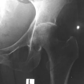
XR
- Soft tissue swelling
- Narrow joint space if > 1/52
- Later subchondral osteoporosis
- End-stage bone destruction seen
Te99 Scan
- Can differentiate from OM
- Excellent screen
USS
- Zarwin Radiology 1993
- 96 patients with painful hips
- None of 40 infected hips had normal USS
Neonate Hip
- Wide joint space ± Subluxed on XR
- Primary OM in metaphysis
- Metaphyseal rarefaction
- Periosteal new bone formation
- ± Erosion of proximal epiphysis
- Fluid in joint on USS
- Te Scan differentiate focal metaphyseal OM
- Also cold if head dead
Differential Diagnoses
Infants & Child
- Acute OM
- Usually less ↓ ROM
- Can get symptomatic effusion
- 70% both
- Transient Synovitis
- More ROM
- Afebrile
- ESR normal
- JRA
- Gradual onset
- Less systemic features
- More ROM
- Perthes
- Trauma
- Haemophilia
- Cellulitis
Adults
- Gout
- Especially in knee
- Peri-articular punched-out lesions
- Pseudogout
- Meniscal & articular calcium
- RA
Management
Antibiotics
- Start after M/C/S
- Start broad-spectrum bacteriocidal
- Gram stain as guide
- Flucloxacillin & Gentamicin in Adults
- Flucloxacillin & Ceftriaxone 40mg/kg/dose q24h IV in Children
- IVAB until systemic toxicity & local swelling subside & CRP normal
- ~ 2/52
- Then 6/52 oral AB
Drainage
- Surgical emergency
- Arthrotomy or Arthroscopy
- Small capsulectomy
- Washout pus
- ± Synovectomy
- Close skin only
- Splint joint in position of function
- Pain relief
- Mobilise as pain allows
- Traditional to immobilise joint to relieve pain in acute phase of disease
- Salter et al 1981 advocates early passive ROM (via CPM) based on rabbit studies which show that this
- Improves cartilage nutrition
- Stimulates chondrocytes to synthesize matrix components
- Prevents adhesions
- Helps clear lysosomal enzymes & purulent exudate
Prognosis
- Worse if
- Treatment delayed (most important)
- Hip
- Associated OM
- Staphylococcus aureus or multiple organism
- Neonate as diagnosis delayed & usually hip
Outcomes of Septic Arthritis (of Hip)
- Normal
- Growth disturbance
- Coxa magna
- Coxa breva
- LLD
- Subluxation
- Dislocation
- Destroyed epiphysis
- Secondary Osteoarthritis
- Ankylosis
- “Tom Smith arthritis”
- Arthritis of the hip following neonatal septic arthritis
- From umbilical manipulation?
- Due to delayed diagnosis
Reviewed by Professor Phong Tran, Head of Orthopaedic Surgery, Western Health. Last updated 10 October 2026.