
- AKA Osteitis Deformans described by Sir James Paget 1877
Definition
- Disorder of bone turnover & remodelling of unknown aetiology
- Disturbance of rate of bone turnover
- 1. OsteoLytic phase (Osteoclastic)
- 2. OsteoSclerotic phase (Osteoblastic)
- 3. Burnt out phase
- Polyostotic or monostotic
- Most asymptomatic
- Incidental XR finding
Epidemiology
- mostly > 50 years old
- 5% of people > 50
- 8% of people > 80
- M3 > F2
- Geographic variations
- wide geographic variation between countries & even cities, e.g. foci of Paget’s in Lancashire & in Malta
- 10-15% of elderly patients in Northern Europe
- virtually non-existent in Japan
- Temporal clusters
- Familial clustering
- may be autosomal dominant inheritance
- relative risk in 1st degree relatives is 7
- Family History 15%
- Most common sites
- Spine
- Femur
- Skull
- Pelvis
Aetiology
- Unknown -? disorder of osteoclasts
- Recent evidence Paramyxovirus
- Measles
- Respiratory Syncytial Virus
- Probably, Pagets disease is caused by infection with a common & widespread virus superimposed on genetic variation for susceptibility
- Viral
- slow virus
- Virus intra-nuclear inclusion bodies in osteoclasts
- measles (paramyxovirus) or adeno-type
- Has never been grown
- Genetic
- A candidate gene on chromosome 18q
- familial clustering
- positive family history in 15% to 30% of patients
- Human leukocyte antigen (HLA) patterns
- Viral
- Associations
- primary hyperparathyroidism
- DISH (14-30%)
- gout & pseudogout
Pathophysiology
- Two Phases (classically divided into 3 parts)
- Active
- Osteolytic (osteoclastic)
- Starts at one end of long bone
- Osteoclasts hyperactive
- Leads to ↑ bone resorption / destruction with
- Osteoporosis
- Fibrovascular hyperplasia
- Resorption stimulates osteoblastic activity
- Woven bone produced rapidly absorbed on both endosteal & periosteal surfaces
- excessive & pathologic (disorganised immature) bone formation by osteoblasts
- Bone soft
- Osteosclerotic (osteoblastic)
- Osteoblastic activity > osteoclastic activity
- Sclerotic ivory-hard bone produced
- abnormal bone remodelling
- Osteolytic (osteoclastic)
- Inactive (“burnt out”)
- Bone remodelling & turnover decreases to normal
- Remaining bone is
- Enlarged
- Brittle
- Sclerotic
- Deformed
- Active
Pathology
Gross
- Distorted bone
- Bowed because structurally weak
- Spongy bone with generalised enlargement
- Thick cortices
- Coarse trabeculae
- Immature woven appearance
Histology
- Irregular segments of mature (lamellar) bone with ↑ cells
- Osteoclasts, Osteoblasts, Fibroblasts
- Osteoclasts
- activity
- number & size
- Nuclei are numerous & remarkably large
- bone turnover is greatly ↑ (up to 40 times normal)
- activity
- Osteoblasts
- activity
- Peritrabecular fibrosis
- also seen in other hypermetabolic conditions such as hyperparathyroidism
- Hypervascular
- Cortex
- thickened
- Corticomedullary demarcation lost
- Trabeculae
- both thick & thin
- Mosaic pattern
- “Burned-out” sclerotic pagetic bone
- 1. Osteolytic phase
- X-ray
- an advancing wedge of osseous rarefaction
- Marked osteoclastic resorption
- Multinucleated osteoclasts with nuclear inclusion bodies line the trabeculae
- Extremely vascular fibrous tissue fills the marrow spaces (osteoporosis circumscripta)
- Inflammatory cells absent
- Cancellous bone
- Trabeculae slender & sparse
- Cortical bone
- Large resorption cavities seen
- Prominent osteoblasts occur concurrently
- Appositional new bone formation
- Woven bone
- Both ↑ osteoclastic & osteoblastic activity seen on the same trabeculae
- Process occurs on both endosteal & periosteal surfaces ® bone ↑ in thickness but is structurally weak ® deformity
- Frenetic cell activity difficult to differentiate from hyperparathyroidism (osteitis fibrosa cystica)
- X-ray
- 2. Osteoblastic phase
- New bone formation predominates over resorption
- Trabeculae are broad & the cortical bone thickens with sclerosis
- Neither cortical nor cancellous in architecture
- No abnormality of mineralisation but may see wide osteoid seams
- Widened lamellae & disorganised cement lines
- Gives diagnostic “Mosaic Pattern”
- Alteration in architecture together with ↑ cement lines leads to structural weakness & facilitates propagation of cracks
- Normal fatty or haematopoietic marrow replaced with fibrovascular connective tissue
- 3. “Burnt-out” phase
- Gradually osteoclastic activity abates & the eroded areas fill with new lamellar bone leaving an irregular pattern of cement lines these mark the limits of the old resorption cavities ® marbled or mosaic appearance on microscopy
- In the late osteoblastic stage the thickened bone becomes increasingly sclerotic & brittle, bone marrow reverts to fat & lining cells on the trabeculae are inactive
- All three phases of the disease may be evident in the same specimen
- Cell activity less intense & vascularity diminished
- Rate of bone turnover not much greater than normal
- Bones enlarged & sclerotic with deformity
- Marrow may appear relatively normal
- 4. ?sarcoma
- Some investigators would add a 4th phase to Paget’s disease: malignant degeneration into a sarcoma
- Patients with Pagets disease have a 30 fold ↑ in the incidence of bone sarcoma (Price 1962)
Clinical Features
- Usually asymptomatic or pain & deformity
- Monostotic in 17% & polyostotic in 83%
- pelvis 70%
- Lumbar spine 50%
- femur 50%
- skull 45%
- tibia 30%
- humerus 30%
- clavicle 13%
- hand & foot 3 – 5%)
- Asymptomatic (incidental finding)
- 20% asymptomatic
- diagnosed from XR taken post trauma
- alkaline phosphatase
- 20% asymptomatic
- Pain in Pagetoid bone
- » “BANISH”
- Bone pain
- Deep, constant & aching
- Due to
- metabolic activity
- periosteal stretching
- vertebra compression Fracture
- If worse at night
- ? sarcoma, impending Fracture
- Arthritis
- Due to
- Abnormal subchondral bone
- Abnormal biomechanics due to deformity
- Usually Medial Hip Osteoarthritis
- Cf. Superior pole with primary Osteoarthritis
- Abnormal subchondral bone
- Due to
- Neurological impingement
- Stenosis
- Central or Lateral Recess
- Cranial Nerve
- Stenosis
- Impending fracture
- Pain on weight bearing
- Sarcoma
- Severe night pain
- Hypercalcaemia
- Bone pain
- » “BANISH”
- Deformity
- Characterised by
- Increased size & abnormal shape of bones
- Thicker cortex
- Bow along stress lines
- Femora bow anterior & lateral
- Tibia bow anterior & lateral – Saber Shin
- Skull enlargement
- Occiput & frontal areas
- Hats don’t fit
- Thoracic Kyphosis
- Arthrokatadysis
- Increased size & abnormal shape of bones
- Characterised by
- Pathological Fracture
- Incidence of 10%
- Most frequent in
- Femoral neck
- Subtrochanteric femur
- Tibia
- Usually transverse
- On convex side (cf. Looser zones in osteomalacia)
- Tension side of bone may have painful stress fractures
- This fracture usually begins as a transverse line on the convexity or tension side of the bone & progresses across the cement lines. The transverse radiolucent line is described as a pseudofracture
- Patients with pseudofracture are usually managed with protected weight bearing & immobilisation in a cast until there is relief of pain. Prophylactic nailing is made problematic by the associated deformity
- “Fracture healing can be impaired, resulting in delayed union & nonunion” (Dee 1997)
- Neurological Compromise
- Cranial Nerve entrapment in foramina
- Neurosensory & Conductive deafness
- Cochlear compressed & ossicle ankylosis both lead to deafness
- 50% of patients deaf
- Softening & basilar invagination with vascular compression
- Brain stem or Cerebellar compression
- Blockage CSF flow with hydrocephalus
- Spinal Stenosis may require surgical decompression
- CVS Compromise
- High-output cardiac failure
- Steal syndrome
- Shunting of blood can cause cerebral hypoxia & spinal cord ischaemia
- Malignant Change
- 1%
- More common in polyostotic form (5%)
- Men twice as likely as women to see malignant change
- 70yo
- 30% bone sarcomas secondary to Paget’s
- Osteosarcoma followed by MFH most common forms
- Survival for Enneking IIB is 15%
- 70yo
- Suspect if previously affected bone painful, swollen & tender
- Large rise in Alk Phos level often seen
- skin temperature
- due to vascularity of bone
Laboratory Investigations
- Uses
- Confirm diagnosis
- Monitor treatment
- CaPO4 & PTH are normal
- Occasionally hypercalcaemic after immobilise
- Alkaline Phosphatase (ALP)
- Hallmark of active disease
Up to 20-30 Times Normal
- 10% of patients with Pagets will have levels within the normal range
- Located in the plasma membrane of osteoblasts
- Reflects
- bone formation
- number & functional state of osteoblasts; that is the level of bone formation
- Correlates roughly with the extent of skeletal involvement
- Serial determinations provide biochemical index of disease activity
- measure annually to monitor disease
- Measure of osteoblastic activity
- Dependent on extent of disease
- May be normal with localised Paget’s
- Rapid ↑ may indicate sarcomatous change
- 50% of ALP is of hepatic origin
- Is There Now Bone Specific ALP?
- Can distinguish Hepatic cause by
- Assessment of other LFT
- Urine Hydroxyproline
- Urinary Hydroxyproline
- Marker of osteoclastic activity
- Reflects collagen turnover
- 24hr urine level measured
- Disadvantage lack of sensitivity
- Useful for extent & progress of disease
- Rapidly reflects response to treatment
- Pyridinium Crosslinks
- New measure of bone resorption
- Measures urinary excretion
- Of hydroxypyridinium crosslinks of collagen
- Derived from degradation of mature bone collagen
- More specific marker than Hydroxyproline
- Normal
- Acid phosphatase
- Serum calcium & phosphate
- Maybe
- Uric acid
- LFTs
- should be done concurrently; if they are elevated then the bony isoenzyme can be isolated
- Urine
- Hydroxyproline
- reflection of osteoclasts function
- not done routinely
- requires a meat-free diet & a 24-hour urine collection
- urinary hydroxy proline to creatinine ratio correlating well with disease activity
- Pyridinoline cross links
- can also be measured in the urine
- N-telopeptides
- are another urinary measure of bone resorption. Crosslaps can be measured in the serum & provide a measure of bone resorption
- Hydroxyproline
- Osteitis Fibrosa cystica
- Fibrous Dysplasia
- Osteoblastic secondaries
- Osteopetrosis
- Hyperparathyroidism
- Lymphoma
Radiology
XR
- Focal bone resorption & formation
- Radiolucencies
- Radiodensities
- Overall bone size enlarged
- Coarse trabecular pattern
Active Stage
- Skull
- Sharp radiolucent areas (Osteoporosis Circumscripta)
- Long bones
- Flame-shaped osteolytic front extending from ends (Flame Sign)
- “flame” advances along the bone
- 1cm/year in untreated patients
- “flame” advances along the bone
- Flame-shaped osteolytic front extending from ends (Flame Sign)
Inactive Stage
- Skull
- Fluffy thick sclerotic bone (Cotton Wool Skull)
- Spine
- Cortical Sclerosis (Picture Frame Vertebra)
- Uniform sclerosis with ivory vertebra (Ivory Body)
- size of the vertebra
- Long bones
- Sclerosis with cortical thickening & coarse trabeculae
- Widened deformed bone
- Coxa Vara
- Anterolateral bowing
- Chalk Stick
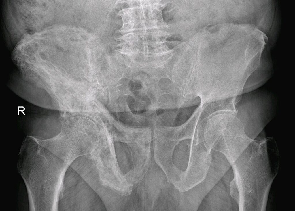
- Pelvis
- Patchy osteolytic/ osteoblastic changes
- Thickening of pelvic brim & iliopectineal line (Brim Sign)
- Protrusio
- Secondary Osteoarthritis
Bone Scan
- All patients at time of Diagnosis to define involved bones
- Can be used to distinguish between areas of tumour & Paget’s
- Only 65% of the lesions seen on bone scan will be seen on X-Rays
- High turnover/ active phase
- May show markedly ↑ uptake in the area
- With treatment & inactive (burnt out) phase
- May be less hot or cold
- Useful
- Confirm diagnosis
- As baseline
- Cold in Hot » Consider Pagetoid Sarcoma
- Gallium scans
- hot with tumours but not with Paget’s
CT Scan
- Diagnose or exclude sarcomatous change
MRI
- Shows new cortical destruction
- Soft tissue mass
- Spinal Stenosis
Management
- Most patients never need treatment
- All patients
- Stage the disease
- full body bone scan
- XR of affected regions
- Stage the disease
- No treatment
- Most
- Asymptomatic patients with near normal body chemistry whose weight bearing bones are not involved
Indications
- Medical Treatment
- Pain
- Neurological complications
- Spinal Stenosis
- Deafness
- Repeated fractures
- Deformity
- Mild Osteoarthritis
- Before & after bone surgery
- Hypercalcaemia due to immobilisation
- High-output cardiac failure
- Surgery
- Severe Osteoarthritis
- Severe malalignment pain
- Pathological fracture
- Sarcomatous degeneration
- Neurosurgery
- Stem or cord compression
Medical Treatment
- Indication is mainly pain
- Aim is to retard osteoclasts
- Hence works best in active disease
- Simple Analgesia
- Use in normal turnover & mild pain
- Calcitonin
- Small polypeptide hormone from the parafollicular cells of thyroid
- Potent inhibitor of osteoclastic activity (direct inactivation of osteoclasts)
- Salmon form most potent
- Expensive
- Nasal Spray or IVI
- Start with 100u daily
- Reduce to weekly after response
- If no response in 3/12 stop
- N & V
- Relapse 30% of patients
- 10% have antibody-mediated resistance
- » Use human form
- Reserve for
- Severe pain due to rapid turnover
- Impending fracture, fracture & post ORIF
- Ie. Diphosphonates contraindicated
- Diphosphonates
- Pyrophosphate analogue
- Decouples osteoblast-osteoclast interaction
- Inhibit normal bone resorption & mineralisation
- With low dose inhibition of bone resorption predominates
- with chronic use or high dose inhibition of mineralisation predominates
- Thus can cause focal osteomalacia
- May lead to pathological fracture
- Thus can cause focal osteomalacia
- » Contraindicated if impending fracture or fracture & postoperative (due to risk of nonunion)
- Use in high turnover disease with pain
- Advantages
- Oral
- Cheaper
- Less relapse than Calcitonin
Etidronate
- Older form
More Risk Osteomalacia
- Etidronate impairs mineralisation at the doses used for Paget’s disease
- Prolonged or high dose courses cause osteomalacia and raise fracture risk
- Courses limited to 5 mg/kg/day for up to six months
- Superseded by more potent bisphosphonates such as zoledronic acid
Alendronate
- Newest
- Least Osteoblasts effects
- Empty stomach
- Upright 1/2 hr
- Pamidronate
- Given IV
- Used by RBH
Serial Management
- Alternate Calcitonin & Bisphosphonate (Alendronate)
- Enhances effects
- Decreases side effects
- Try medical treatment first with spinal stenosis
- May ↓ ‘steal’ syndrome
- Mithramycin
- Antibiotic with cytotoxic properties
- Potent osteoclast inhibitor
- Rapidly relieves pain from Pagets
- Causes hypocalcaemia
- Main indication due to severe side-effects is Pagets Paraplegia
- Surgery
- Stress Fractures
- Try bracing & NWB
- Prophylactic ORIF if not healed 3/12
- Earlier if NOF fracture on tension side
- Stress Fractures
- Complete fracture
- Most commonly occur in
- Femur
- Tibia
- Humerus
- Radius
- Ulna
- Fracture may be the first sign of Paget’s disease in 2/3 of patients
- Classically transverse orientation with disruption of the periosteal sleeve & comminution
- Most fractures will heal with abundant callus
- Non-union common
- Correct deformity prior ORIF
- May require osteotomy for femoral shaft or subtrochanteric fracture
- Femoral neck fracture
- Subcapital/ Transcervical
- Hemiarthroplasty preferable
- Almost 100% non-union with internal fixation
- If protrusio consider THR
- Subtrochanteric/ Intertrochanteric
- More common
- Tend to unite after internal fixation
- Higher non-union with Subtrochanteric
- Higher nonunion seen in
- Subtrochanteric fracture
- Sclerotic phase
- Usually heal with callus +++
- Calcitonin useful to promote healing
- Subcapital/ Transcervical
- Correct deformity prior ORIF
- Most commonly occur in
- Varus deformity of shaft will make Intramedullary nailing difficult & may require osteotomy
- Arthritis
- TJR – Good success rates
- Slight ↑ risk loosening
- Pagets & TJA
- No difference from other primary THR if cemented with respect to aseptic loosening
- No studies for TKR
- Always correct proximal deformity first (eg hip if knee affected)
- Total Hip Arthroplasty
- Similar indications as non-Pagetoid disease
- Preoperative
- Need to ensure that pain from the joint disease & not bone disease
- Painful joint with Pagets
- Bone pain with active Pagets
- Insufficiency Fracture
- Osteosarcoma
- Arthritis
- Neurological compression
- Proximal problem
- Eg. with painful knee consider hip or spine
- Painful joint with Pagets
- Essential to get long leg, weight-bearing films
- ALP
- If ALP > 700 then need to control medically prior to OT
- » Increased bleeding
- » Catastrophic hypercalcaemia
- Endocrinology Review
- Anaesthetic review
- Need to ensure that pain from the joint disease & not bone disease
- Intraoperative
- Deformity
- Coxa Vara
- » Tendency varize stem
- May need femoral osteotomy to allow insertion of stem
- Always get full length films
- » Tendency varize stem
- Protrusio Acetabuli
- Coxa Vara
- Increased bleeding
- Cross match blood
- Sclerotic
- Difficult reaming
- Sharp reamers
- Difficult reaming
- Deformity
- Postoperative
- HO
- 52% vs 5 % in normal patients (Merkow et al 1984)
- Treat with NSAID
- Early loosening
- Not substantiated (Halliday says similar results to primary THR in non-Pagetoid bone)
- HO
- Osteotomy
- Performed to
- Improve deformity
- Improve mechanics of weight bearing joint
- Good results with HTO
- Intertrochanteric hip osteotomy less reliable
- Slow healing if sclerotic phase
- Hugh English advises strongly against this…
- Not advised to do osteotomy » fails to heal » high nonunion rate
- Performed to
- Spinal Stenosis
- Middle-aged man with increasing paresis over a year
- Usually multiple levels
Principles
- There are three principal classes of drugs all are primarily inhibitors of bone resorption
- each induces a rapid fall in hydroxy proline values within the first few days of treatment & this is followed by a later fall in alkaline phosphatase
- Disease monitoring with Alkaline phosphatase levels
- normal = 30-120 units/L (analytical error 9u/L)
- Course of therapy usually continues six months, & 60% achieve remission without relapse for 5 years
- If relapse occurs
- recommence therapy
- For surgery should commence 3 months prior to surgery & continue for six weeks post surgery
Bisphosphonates
- Action
- Potent inhibitors of bone resorption
- Bind to hydroxyapatite crystals – poisons osteoclasts; induces apoptosis & decreases their recruitment
- New bone formed during treatment is lamellar
- goal of treatment i
- to achieve / maintain a mid-range normal level of SAP
- Treatment is restarted when the SAP climbs to 25% higher than this
- Agents
- alendronate (Fosamax)
- agent of choice
- orally
- doesn’t cause osteomalacia
- more effective than etidronate or calcitonin
- 40mg orally daily for six months
- Compare with dose for osteoporosis (10mg/day)
- Side effects
- oesophagitis
- patient should remain upright for 30mins after taking the tablet
- oesophagitis
- Pamidronate (Aredia)
- intravenously
- 60mg over four hours every 8-12 months
- Side effects
- iridocyclitis or a flu like illness
Calcitonin
- Obsolete now
- Binds to adenylate cyclase & turns off osteoclasts
- Dosage: 50-100U intranasally daily then three times weekly
- Causes fall in serum calcium & ↓ resorption of bone
- Urinary hydroxyproline decreases in a matter of days
- Long term benefits
- -Relief of bone pain – occurs in a couple of weeks
- -Reduction in cardiac output
- -May reverse neurological deficits
- -Healing of osteolytic lesions
- -Reduction of bleeding associated with surgery
- Side effects
- -Nausea & flushing (up to 30%)
- -Diarrhoea
- -Pain at injection site
- -Resistance can develop (up to 20% in patients treated with salmon calcitonin)
- Treatment needs to be continued long term
Orthopaedic Surgical Procedures
Fractures
- most common complication
- 10% of patients with extensive Paget’s disease will suffer pathologic fracture, usually of the femur, tibia, or forearm
- High complication rate
- Delayed union or non-union
- 15-40% nonunion
-
- sclerotic phase of the disease
- subtroch femurs
- Delayed union or non-union
- Often fracture through sarcoma
- 5-20% of pagetoid fractures!
- Stress or pseudofractures
- Commonly
- tibia
- proximal femur
- convex side of bone
- Treatment
- protected weight bearing
- If pain persists for more than 3-6 months prophylactic surgical treatment is warranted
- Commonly
- Completed fractures
- Diaphyseal
- transverse or short oblique – chalk stick
- Treatment
- IM
- Passage of an intramedullary device may be technically difficult because of the disordered architecture of the pagetic bone
- IM
- Femoral neck fractures
- Subcapitals
- nonunion rate of 75-90%
- Low threshold for hemiarthroplasty
- Intertrochanteric fractures
- will usually unite
- Subcapitals
- With these exceptions, fractures in Paget’s disease tend to heal with abundant callus
- Delayed union or nonunion is more common in the sclerotic burned-out phase of the disease. Even after successful union, protective bracing may be needed for 3–6 months to allow for remodelling, which is slow
- Diaphyseal
- Corrective osteotomies are frequently required
Elective Orthopaedics
Hip Replacement
- Preoperative
- Referred pain
- ? cause of pain hip vs spine
- many patients also have involvement of the lumbar spine
- carefully separate possible lumbar spine pain from hip pain before undertaking arthroplasty of the hip
- Diagnostic block should be considered
- Medical referral
- Bisphosphonates
- Not in active phase
- Referred pain
- Intraoperative
- Pelvic
- 25% rate of protrusio
- Femoral
- Varus deformity
- Anterolateral bowing of the femur (usually not a major problem because the femoral canal is widened)
- Distorted medullary canal
- Prosthesis
- Uncemented preferred
- Cement should not be used
- Because of active remodelling
- Cement should not be used
- Uncemented preferred
- Increased blood loss
- should pre treat with bisphosphonates
- Difficulty in reaming sclerotic bone
- Pelvic
- Postoperative
- Rate of loosening is ↑ (10-15% at 10 years)
- Heterotopic ossification – up to 50%
Reviewed by Professor Phong Tran, Head of Orthopaedic Surgery, Western Health. Last updated 10 October 2026.