
- Four forms
- different consequences
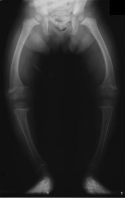
- Predictable by direction of bow
- Lateral bowing
- normal variant during 1st yr of life
- usually resolves spontaneously
- Anteromedial Bowing
- usually associated with fibular hemimelia
- associated with short tibia
- May require leg lengthening
- kyphosis
- Posteromedial Bowing
- presents at birth with shortened limb & grossly calcaneus foot
- Foot often lies with dorsum against anterior leg
- usually benign packaging defect
- Probably caused by intrauterine fracture or malposition
- improves in first two years
- often confused with calcaneoValgus foot
- nothing to do with Neurofibromatosis
- usually remodels
- may need correction of residual deformity and most need treatment of LLD > either leg lengthening or physeodesis
- Foot position improves in advance of bowing
- Tibia & fibula intact both bowed
- Bow most pronounced in distal half of each
- Commonly both are short = problem
- Proportional differences in lengths remain stable
- if severe after 3 yrs > osteotomy or epiphysiodesis?
- associated with
- Calcaneus foot
- Triceps surae weakness
- Extension contracture of ankle
- Anisomelia
- Anterolateral Bowing
- Dangerous form
- associated with pseudarthrosis of tibia
- Prevent fracture by bracing
- also seen with tibial hemimelia
- indicative of Congenital pseudarthrosis
- rare Congenital type with duplication of first ray associated anteroLateral bow
- Surgery
- very difficult
- if severely dysplastic prob need some form of rod
Reviewed by Professor Phong Tran, Head of Orthopaedic Surgery, Western Health. Last updated 10 October 2026.