
Definition
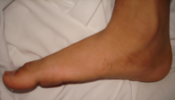
- Loss of medial longitudinal arch with hindfoot valgus and forefoot abduction
- Talar head plantarflexed and uncovered medially
Aetiology
- Flexible idiopathic flatfoot
- Neuromuscular causes are spastic CP, myelomeningocele and muscular dystrophy
- Rigid causes are tarsal coalition and congenital vertical talus
- In CP, spastic peronei and gastrocsoleus overpower a weak tibialis posterior
Epidemiology
- Planovalgus is the most common foot deformity in spastic diplegia and quadriplegia
Clinical Features
- Hindfoot valgus and too many toes sign
- Arch reconstitutes on tiptoe and Jack test in the flexible foot
- Callus and pressure under the talar head
- Lever arm dysfunction weakens push off in CP gait
- Silfverskiöld test for gastrocnemius contracture
- Assess tone, selective motor control and walking ability (GMFCS)
Investigations
- Standing AP and lateral foot radiographs
- Lateral talar first metatarsal (Meary) angle apex plantar
- Reduced talonavicular coverage on AP view
- Standing ankle AP to exclude ankle valgus
Management
- Asymptomatic flexible idiopathic flatfoot needs no treatment
- SMO or AFO in neuromuscular feet
- Botulinum toxin and casting for dynamic equinus in CP
- Gastrocnemius recession or tendo Achillis lengthening for fixed equinus
- Calcaneal lengthening osteotomy (Evans) for flexible deformity in ambulant children
- Extra articular subtalar arthrodesis (Grice) in younger children with severe neuromuscular deformity
- Subtalar or triple arthrodesis for rigid or severe deformity in older children
- Medial malleolar screw hemiepiphysiodesis for coexisting ankle valgus
Complications
- Undercorrection or recurrence, more common in neuromuscular feet
- Calcaneocuboid subluxation after calcaneal lengthening
- Adjacent joint arthritis after arthrodesis
Reviewed by Professor Phong Tran, Head of Orthopaedic Surgery, Western Health. Last updated 10 October 2026.