
Indications
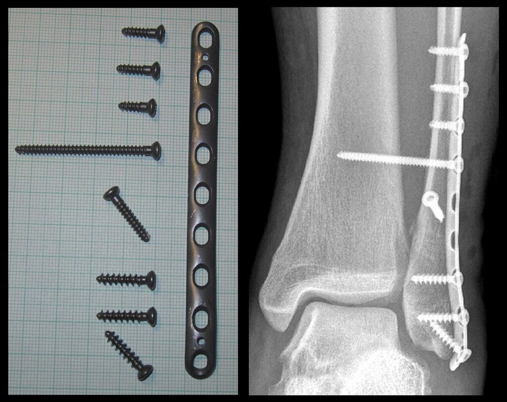
- Talar shift or medial clear space widening
- Bimalleolar and trimalleolar fractures
- Syndesmotic instability
- Weber B fractures with an incompetent deltoid ligament
- Isolated Weber B fractures with a stable mortise on stress or weight bearing views are treated in a boot
Position
- Supine, bump under the ipsilateral hip, thigh tourniquet
- Image intensifier from the opposite side
Operative Steps
- Lateral approach over the fibula, protecting the superficial peroneal nerve
- Lag screw and neutralisation plate, or posterolateral antiglide plate
- Medial malleolus with two partially threaded cancellous screws perpendicular to the fracture
- Fix large or displaced posterior malleolar fragments through a posterolateral approach
- Stress the syndesmosis with the Cotton test or external rotation stress
- Fix syndesmotic instability with screws or suture button under direct or fluoroscopic reduction
- Restore fibular length and rotation, checked with the dime sign and talocrural angle
- Fix the syndesmosis with the ankle in neutral
Postoperative Care
- Early weight bearing in a boot when fixation is stable
- Diabetic and neuropathic patients need longer protection
- Radiographs at 2 and 6 weeks
- Hardware removal only if symptomatic
Complications
- Wound breakdown, worse in diabetes and smokers
- Syndesmotic malreduction
- Superficial peroneal nerve injury
- Symptomatic hardware
- Wound breakdown, higher in smokers and diabetics
- Malreduction of the syndesmosis
- Post-traumatic arthritis
Related Pages
Reviewed by Professor Phong Tran, Head of Orthopaedic Surgery, Western Health. Last updated 10 October 2026.