
Indications
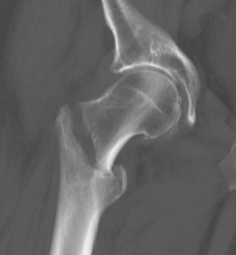
- Stable intertrochanteric fractures with a sliding hip screw
- Unstable patterns, reverse obliquity and subtrochanteric extension with a cephalomedullary nail
- AO 31A1 fractures are stable, A2 and A3 are unstable
- Lateral wall thickness under about 20 mm predicts failure with a sliding hip screw
Position
- Supine on a traction table, contralateral leg in a well leg holder or scissor position
- Image intensifier between the legs
- Pad the perineal post to avoid pudendal nerve injury
Operative Steps
- Closed reduction with traction and internal rotation, confirmed in AP and lateral views
- Guide wire centre-centre in the head on both views
- Tip apex distance under 25 mm to reduce cut out (Baumgaertner)
- Ream, insert lag screw, then side plate or nail
- Compress with the screw before locking
- Distal locking for long nails or length unstable fractures
- Reduce before reaming, open reduction with clamps if closed reduction fails
- Avoid varus and posterior sag of the neck
- Check rotation against the other leg
Postoperative Care
- Weight bear as tolerated
- Check for varus collapse at 6 weeks
- Orthogeriatric care and delirium screening
- Osteoporosis treatment
Complications
- Cut out, most common mechanical failure
- Varus malunion and excess shortening
- Periprosthetic fracture at the nail tip
- Lateral thigh pain from prominent hardware
- Infection
- Medial perforation of the lag screw (Z effect) with two screw nails
Related Pages
Reviewed by Professor Phong Tran, Head of Orthopaedic Surgery, Western Health. Last updated 10 October 2026.