
Aetiology
- Muscle imbalance acting on a growing foot produces progressive deformity
- Upper motor neuron causes are CP and spinal cord lesions
- Lower motor neuron causes are myelomeningocele and polio
- Peripheral nerve cause is CMT
- Myopathic cause is DMD
Clinical Features
- Equinus is the most common deformity
- Planovalgus in diplegia and quadriplegia
- Equinovarus in hemiplegia from tibialis posterior or tibialis anterior overactivity
Spina bifida
- Deformity depends on neurological level
- Rigid equinovarus (teratologic clubfoot) common
- Calcaneus deformity in low lumbar levels from unopposed tibialis anterior
- Insensate feet at risk of pressure ulcers
CMT
- Cavovarus from weak tibialis anterior and peroneus brevis
- Strong peroneus longus and tibialis posterior drive the deformity
- Claw toes from intrinsic weakness
DMD
- Equinovarus from tight gastrocsoleus and tibialis posterior
Investigations
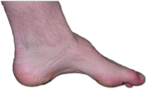
- Standing foot radiographs
- Coleman block test for hindfoot flexibility in cavovarus
- MRI spine for unilateral or unexplained deformity
- Nerve conduction and genetic testing (PMP22 duplication in CMT1A)
- Gait analysis in CP before multilevel surgery
Management
- Aim for a plantigrade, braceable and shoeable foot
- Physiotherapy, AFO and botulinum toxin in CP
- Tendon transfer for dynamic deformity with adequate strength
- Split tibialis posterior or split tibialis anterior transfer for varus in hemiplegia
- Simple tenotomy favoured in spina bifida as transferred muscle function is unpredictable
- Osteotomy for fixed bony deformity
- Arthrodesis as salvage, avoided in insensate feet
Reviewed by Professor Phong Tran, Head of Orthopaedic Surgery, Western Health. Last updated 10 October 2026.