
- Most common accessory bone of the foot
- Located at Medial plantar border of the navicular
- Associated with Tibialis Posterior tendon
- Accessory ossicles are derived from unfused ossification centres
- Synonyms: Os tibiale; Os tibiale externum; Naviculare secundum
Incidence
- 21% incidence; 89% of cases are bilateral
- One of the most common accessory ossicles in the foot It is seen over the medial pole of the navicular bone, usually in adolescent patients
- It is most commonly symptomatic in the 2nd decade of life and causes medial foot pain
- <1% of patients become symptomatic.
- Usually affects teens and young adults
- More frequent in females
Aetiology
- variant of normal anatomy.
- It may become symptomatic from the bony prominence impinging against shoe wear.
- The patient may have diffuse medial and plantar arch pain.
- It may cause problems by destabilizing the insertion and diminishing the pull of the PTT.
- In patients with associated severe flatfoot deformity, lateral pain may occur secondary to impingement of the calcaneus against the fibula.
- A traumatic event can cause injury to the fibrocartilaginous synchondrosis that attaches the ossicle to the main navicular.
Associated Conditions
- Flatfoot deformity
- Secondary Achilles tendon contracture
Symptoms
- Considered an incidental finding on radiographs, but may become symptomatic
- bursa, redness, irritation, local tenderness
- Often presents in adolescent patients or young adults, with flatfoot deformity and arch pain
Classification
3 major types of accessory navicular adjacent to the posteromedial navicular tuberosity
| Type | Description | Incidence |
|---|---|---|
| I | Small, 2 – 3 mm sesamoid bone in the PTT (os tibiale externum) | ~30% |
| II | Larger ossicle than type I Secondary ossification centre of the navicular bone | ~50% |
| III | Enlarged navicular tuberosity (cornate navicular) considered a fused variant of a type II, often with pointed shape | ~20% |
X-ray
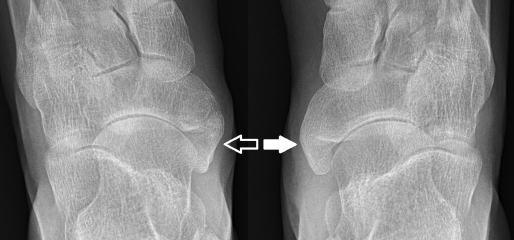
- AP, Lat & 45 degree eversion oblique
- Navicular is the last tarsal bone to ossify from multiple ossification centres
- Females – 1-3.5 yr
- Male – 3.0- 5.5 yr
- Smooth margins with well-formed cortex differentiate this condition from acute fracture
- There is no evidence that the longitudinal arch is any different, essentially an incidental finding
Bone Scan
- May show increased activity over an accessory navicular
- May be needed if a navicular stress fracture is suspected in the differential diagnosis
MRI
- Useful when plain films are unremarkable
- Often, a type-II accessory navicular is attached to the tuberosity by a fibrocartilage or hyaline cartilage layer, and MRI may show soft-tissue oedema consistent with a synchondrosis sprain or tear.
- Altered signal intensity and bone marrow oedema, suggestive of chronic stress and/or osteonecrosis
- Also helpful in showing PTT degeneration
Differential Diagnosis
- Navicular fracture may mimic an acute avulsion fracture of the tuberosity of the navicular.
- Posterior tibial tendinitis
- Stress fracture of navicular
Treatment
- Most patients are asymptomatic or are successfully managed conservatively
- rest and avoid athletics or aggravating activities.
- Anti-inflammatory medication
- Shoe-wear modification
- use of a softer, wider shoe
- If flatfoot is present, a medial arch support may be useful, but often the patient may not tolerate it because of direct pressure on the ossicle.
- Below-the-knee walking cast or removable fracture boot may be used for 3-6 weeks for persistent symptoms.
- Physiotherapy – strengthening exercises
- Surgical
- Rarely simple surgical excision
- Options
- Kidner procedure, the accessory navicular is excised, and the PTT is rerouted into a more plantar position
- excision of the ossicle and reattachment of the PTT insertion to the navicular, with suture anchors or sutures passed through drill holes
- Options
- Severe flatfoot deformity with lateral impingement symptoms
- may require concomitant osteotomy of the calcaneus and/or medial column of the foot to improve alignment and decrease mechanical stress of the PTT insertion.
- Rarely simple surgical excision
Reviewed by Professor Phong Tran, Head of Orthopaedic Surgery, Western Health. Last updated 10 October 2026.