
Question
A 45 year old woman has an incidental cartilage lesion in her proximal humerus found on a shoulder radiograph. A second patient aged 55 has a painful 7 cm lesion in the proximal femur with endosteal scalloping.
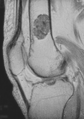
- How do you distinguish enchondroma from chondrosarcoma?
- What investigations would you request?
- How would you manage each patient?
Answer
Enchondroma versus chondrosarcoma
- Enchondromas are usually painless and found incidentally
- Pain not explained by another cause suggests malignancy
- Size over 5 cm and axial or proximal limb site favour chondrosarcoma
- Deep endosteal scalloping over two thirds of the cortex, cortical breach, periosteal reaction and a soft tissue mass favour chondrosarcoma
- Rings and arcs calcification is seen in both
Investigations
- Radiographs and MRI of the whole lesion
- CT for cortical scalloping and destruction
- Biopsy is unreliable for low grade lesions because of sampling error, so imaging and clinical features guide decisions
- Refer suspected chondrosarcoma to a sarcoma centre
Management
- The first patient has a likely enchondroma, managed with repeat radiographs at 6 and 12 months
- The second patient has a likely chondrosarcoma
- Atypical cartilaginous tumour (grade 1) of the long bones can be treated with extended curettage
- Grade 2 and 3 and axial tumours need wide resection
- Chondrosarcoma is resistant to chemotherapy and radiotherapy
- Ollier disease and Maffucci syndrome carry a high risk of malignant change
Related pages
Author Contributions
Orthofracs team
Reviewed by Professor Phong Tran, Head of Orthopaedic Surgery, Western Health. Last updated 10 October 2026.