
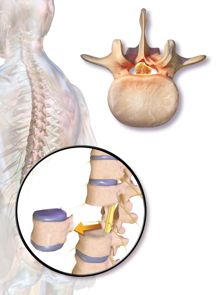
Indications
- Biopsy for suspected spinal tumour, indeterminate lesion, or vertebral osteomyelitis without a culture result
- Vertebrectomy for tumour with cord compression or instability, including solitary metastasis or primary sarcoma
- Unstable burst fracture with canal compromise and neurological deficit
- Vertebral osteomyelitis with abscess, collapse or progressive neurology
- Rigid kyphosis needing vertebral column resection
Position
- Prone for CT guided transpedicular biopsy and posterior vertebrectomy
- Jackson frame with abdomen free for posterior surgery
- Lateral decubitus for thoracotomy and retroperitoneal approaches
Operative Steps
- Plan the biopsy tract with the surgeon who will perform definitive resection, so the tract can be excised en bloc
- Refer suspected primary sarcoma to the treating sarcoma centre before any biopsy
- Use a transpedicular route for vertebral body lesions, which keeps tumour within the pedicle and avoids contaminating the paraspinal space
- Send histology plus aerobic, anaerobic, mycobacterial and fungal cultures
- Take cultures before antibiotics in the stable patient with suspected infection
- Embolise hypervascular tumours, such as renal and thyroid metastases, before resection
- Choose the approach. Posterior transpedicular reaches all levels, anterior gives direct body access, and lateral extracavitary combines both
- Record baseline SSEP and MEP and maintain mean arterial pressure for cord perfusion
- Posterior route. Place pedicle screws two levels above and below, laminectomy, then ligate the thoracic nerve root as needed
- Excise the body piecemeal, remove adjacent discs, and prepare endplates. Protect the dura with a malleable retractor
- Reconstruct with an expandable titanium cage or structural strut graft (fibula or iliac crest), plus cement in tumour
- Contour rods, compress, confirm dural decompression and close over a drain
Postoperative Care
- Neurological observations hourly, with urgent MRI for any new deficit
- Early mobilisation with bracing guided by stability, and venous thromboembolism prophylaxis
- Adjuvant radiotherapy or antimicrobial therapy guided by histology and culture
Complications
- Cord or root injury, with neuromonitoring change as the early warning
- Massive bleeding from epidural veins or hypervascular tumour
- Dural tear and cerebrospinal fluid leak
- Cage subsidence, migration or rod failure
- Pneumothorax after thoracic biopsy, and great vessel or thoracic duct injury in anterior approaches
- Tumour seeding along the biopsy tract, and surgical site infection
Reviewed by Professor Phong Tran, Head of Orthopaedic Surgery, Western Health. Last updated 20 October 2026.