
Question
A 10 year old boy has an incidental lesion in his distal tibia after an ankle sprain. Radiographs are taken over several years.
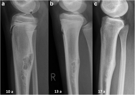
- Describe the radiographs and give the diagnosis.
- When does this lesion need treatment?
- What syndromes are associated with multiple lesions?
Answer
Radiographs
- Eccentric, lobulated, lytic metaphyseal lesion with a sclerotic scalloped border
- Moves away from the physis and ossifies over time
- Non-ossifying fibroma (fibroxanthoma)
- No biopsy needed with classic appearance
Treatment
- Observe, most heal by skeletal maturity
- Pathological fracture through lesions over 50% of the bone width and over 33 mm long (Arata criteria)
- Curettage and bone grafting for large lesions in weight bearing bones or after fracture
- Most fractures heal with casting, then the lesion is treated
Multiple lesions
- Jaffe-Campanacci syndrome, café au lait spots, intellectual disability and multiple NOFs
- Neurofibromatosis type 1
Related pages
Author Contributions
Orthofracs team
Reviewed by Professor Phong Tran, Head of Orthopaedic Surgery, Western Health. Last updated 10 October 2026.